Human ApoCs SDS-PAGE Standard | ABMC-SP4
- SKU:
- ABMC-SP4
- Availability:
- Usually shipped in 5 working days
- Size:
- 0.1 ml
Frequently bought together:
Description
Human ApoCs SDS-PAGE Standard | ABMC-SP4
| Concentration: | 0.20 mg/ml (vortex before apply for gel electrophoresis) |
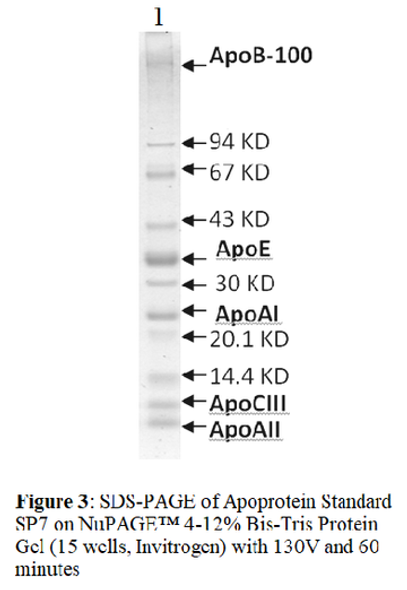
| Size: | 100 μl (20 applications of 5 μl each). ApoCs Standard is a ready-to-use format - no mixing, heating or reducing required. It is pre-reduced and contains reducing reagents in the loading buffer. The protein standards resolve into bands with 6.6 kDa for apoCI, 8.5 kDa for apoCII, and 8.7 kDa for ApoCIII; ApoAII appears as weak visible band between ApoCI and ApoCII. |
|
Source: |
From purified ApoCs and ApoAII; fresh human plasma that has tested negative for Hepatitis C, HIV-I and HIV-II antibodies as well as Hepatitis surface antigens. |
|
Use: |
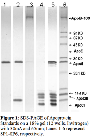
The Un-Stained ApoCs Standard allows you to check the result of your gel run and to judge western transfer efficiency. The attached gel was running with 4 µl of each for lane 1 (ApoCs) and lane 2 (Std Mix2) on an Invitrogen 18% SDS-Gel. |
|
Buffer: |
In loading buffer consists of Tris-HCl, MeSH, SDS, glycerol, and bromophenol. |
|
Storage: |
-20°C for short and long-term storage. Aliquot to avoid repeated freezing and thawing. |
*These products are for research or manufacturing use only, not for use in human therapeutic or diagnostic applications.
IMPORTANCE
ApoCI contains 57 amino acid residues and the m.w. is 6.6 kDa (Jackson et al., 1974). ApoCI has been found to activate LCAT (Liu and Subbaiah 1993).
ApoCII contains 78 amino acid residues. The m.w. is 8.5 kDa (Jackson et al., 1977). The lipid-binding domain of Apo CII is localized to a region in the N-terminal with a strongly amphipathic nature (Macphee et al., 1999).


