DNA Replication Fork
DNA Replication Fork: Labeled Diagram, Function, and Definition
Explore what is a replication fork, see a labeled DNA replication fork diagram, and understand where the replication fork is in the genome. Learn its key role in DNA synthesis.
What Is a Replication Fork?
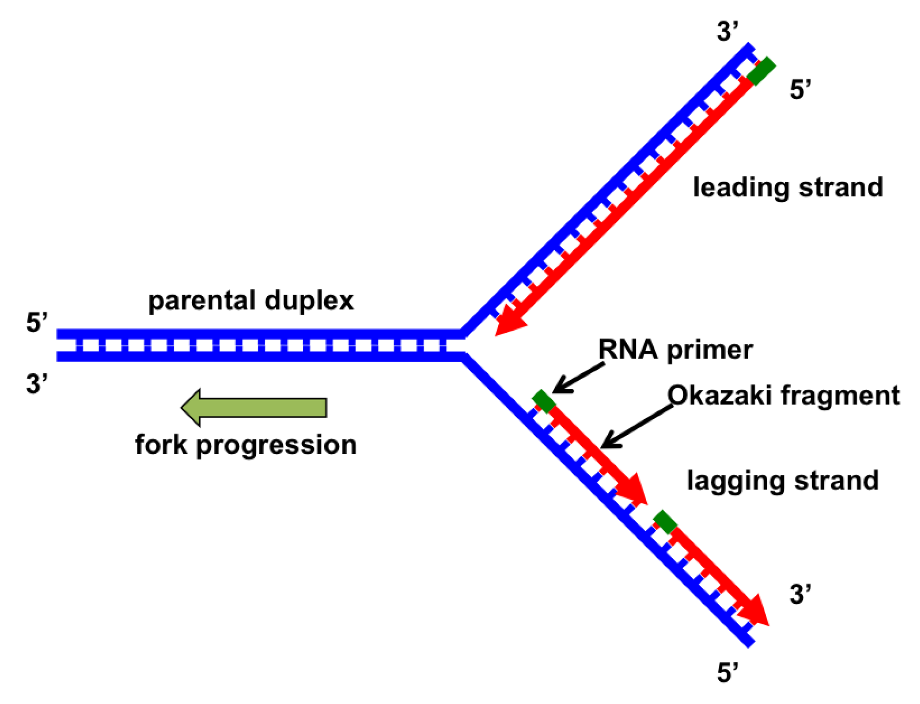
The replication fork is the Y-shaped region of DNA where the double helix unwinds, allowing DNA polymerase to synthesize new complementary strands. This site marks the active area where DNA replication is happening.

replication fork diagram

DNA Replication Fork Structure and Function
At each DNA replication fork, two strands are produced:
- The leading strand is synthesized continuously.
- The lagging strand is synthesized in short fragments called Okazaki fragments.
Multiple enzymes operate here:
- Helicase unwinds the DNA.
- Primase lays RNA primers.
- DNA polymerase extends the new strands.
- Ligase joins lagging strand fragments.
These enzymes work in a coordinated complex that drives semiconservative replication.
Replication Fork Diagram (Labeled)
A replication fork diagram helps visualize how the machinery operates at the molecular level. In a typical DNA replication fork labeled diagram, you will find:
- Helicase at the fork apex.
- Primase ahead of polymerase.
- Leading strand synthesized in the direction of the fork.
- Lagging strand synthesized away from the fork.
- SSB proteins stabilizing unwound DNA.
Where Is the Replication Fork Located?
The replication fork forms at origins of replication, which are specific sequences where the unwinding of DNA begins. In:
- Eukaryotic cells: multiple replication forks form simultaneously along linear chromosomes.
- Prokaryotic cells: typically one origin forms two forks that progress in opposite directions.
DNA Replication Fork Animation
During DNA replication inside a cell, each of the two old DNA strands serves as a template for the formation of an entire new strand. Because each of the to daughters of a dividing cell inherits a new DNA double helix containing one old and one new strand, the DNA double helix is said to be replicated “semiconservetively” by DNA polymerase.
Analyses carried out in the early 1960s on whole replicating chromosomes revealed a localized region of replication that moves progressively along the parental DNA double helix. Because of its Y shaped structure, this reactive region is called a replication fork. At a replication fork, the DNA both new daughter strand is synthesized by a multienzyme complex that contains the DNA polymerase.
Summary Table: DNA Replication Fork Explained
| Term | Description |
|---|---|
| Replication fork | Y-shaped structure formed during DNA unwinding |
| DNA replication fork | Specific region where DNA is duplicated by polymerase |
| Replication fork diagram | Visual representation of replication process and enzyme positions |
| Replication fork labeled | Annotated diagram showing helicase, primase, polymerase, ligase, etc. |
| Where is the replication fork | Found at every origin of DNA synthesis during S-phase |
| Replication forks | Multiple forks occur simultaneously in eukaryotic chromosomes |
There are no products listed under this category.
