
Imaging System
Comprehensive Solutions for Molecular Imaging and Blotting Workflows
Explore ATTO’s innovative line of scientific imaging solutions engineered for protein detection, DNA/RNA visualization, and live cell analysis.
1-Chemiluminescent Detection Imaging Systems
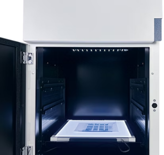
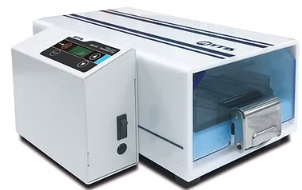
A- WSE-6370 LuminoGraph III Lite

ATTO’s imaging systems are engineered to support diverse laboratory applications, offering accurate visualization and documentation for:
- Western Blot & Dot Blot Imaging – Chemiluminescent or colorimetric detection.
- DNA Gel Electrophoresis – Ethidium bromide, SYBR, GelRed, and other stains.
- Bioluminescence Imaging – Ideal for luciferase assays and reporter gene studies.
- Protein Gel Analysis – Coomassie Blue, silver stain, and other colorimetric methods.
Smart Imaging Solutions Designed for Performance and Usability
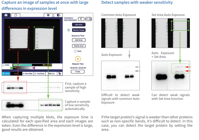
- AutoExposure Mode - Automatically calculates optimal exposure time and captures high-quality images with precision.
- Auto Contrast & Inverse Image Tools - Built-in image analysis software enables real-time contrast adjustment and inverted visualization for enhanced clarity.
- User-Friendly Interface - Simple icon-driven layout for intuitive operation — no steep learning curve.
- Multiple Detection Modes - Choose from auto exposure, summation, and other customizable capture modes.
- User Management & File Organization - Personal folders and user registration for secure, organized image storage and access.
- High-Sensitivity Imaging Hardware - Equipped with a 6-megapixel cooled CCD camera for ultra-sensitive, high-resolution imaging.
- F0.8 Ultra-Fast Lens - High numerical aperture lens improves light capture and sharpness in low-signal conditions.
- CS Analyzer Software (ATTO Original) - Advanced image analysis software with quantitative tools, pattern correction, and band normalization functions.
- Quantitative Band Analysis - Accurately measure band intensity, normalize to housekeeping proteins, and revise distorted band patterns.
- Windows-Compatible with Free Software Updates - Install on standard Windows PCs with continuous access to free software upgrades.
B- WSE-6170 LuminoGraph I CMOS
Versatile Imaging for Chemiluminescence, Fluorescence, and Gel Documentation
The LuminoGraph I CMOS is designed for multi-mode imaging in molecular biology and protein analysis. It enables researchers to:
- Capture chemiluminescent images of Western blots with high sensitivity and clarity.
- Visualize fluorescently labeled gels and proteins (optional module available).
- Perform bright-field imaging of Coomassie Brilliant Blue (CBB)-stained protein gels (optional).
- Quantify sample concentration and molecular weight directly from image data.
- Merge chemiluminescent images with prestained protein marker images for complete band identification.
|
White light source (Optional)
|
Cyan Transparent Light Source (Optional)
|
|
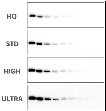
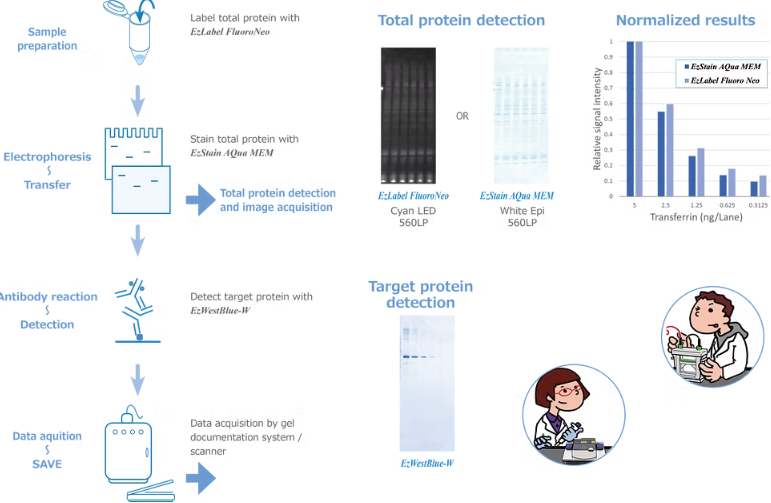
It shows the results of western blotting of transferrin proteins that in a 1/2 dilution sequence from 10ng/lane with anti-transferrin antibodies. It is a photographic image of luminescence after reaction with an HRP luminescence substrate.
|
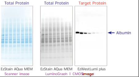
A sample of HeLa cell extraction protein added 0-5 ng of human albumin per lane was separated by u-PAGEL H and transcribed by QBlot Mini, and the total protein on the PVDF membrane was detected by EzStain AQua MEM.
|
2- Western Blotting Solutions
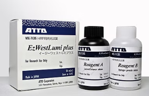
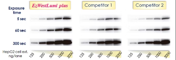
Use for luminescence detection by HRP in western blotting.
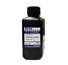
EzWestLumi is a chemiluminescent substrate to detect horseradish peroxidase (HRP) on the western blotting membrane.
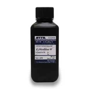
EzWestBlue W is a TMB substrate for horseradish peroxidase (HRP) used for the colorimetric detection of immunoblotting and not applicable to ELISA.

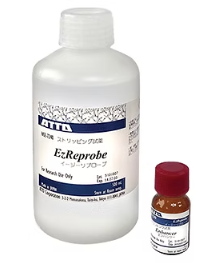
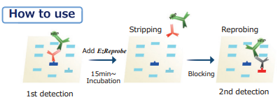
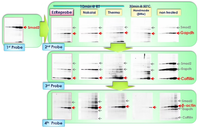
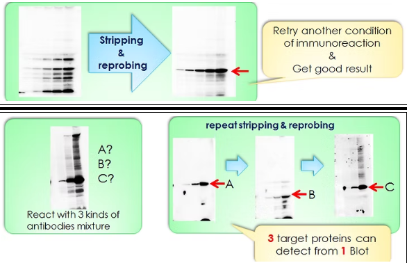
EzReprobe is a stripping reagent to remove the 1st and 2nd antibodies from the membrane after the western blotting reaction and Repeatable stripping and reprobing reaction.
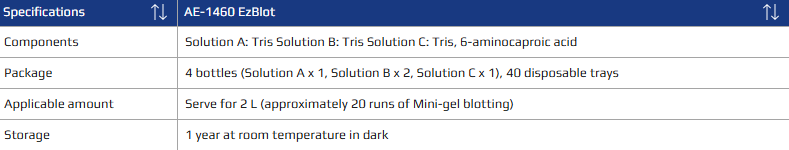
E- AE-1460 EzBlot
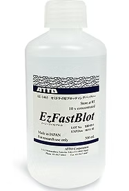
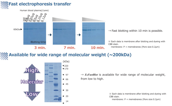
F- AE-1465 EzFastBlot
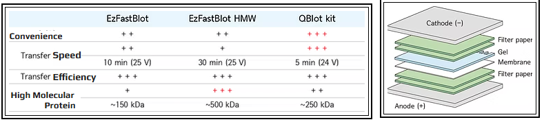
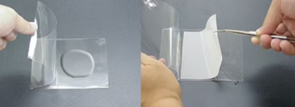
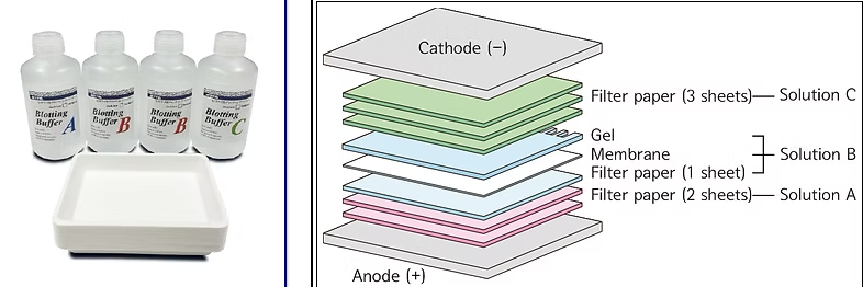
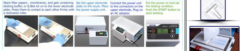
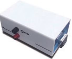
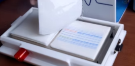
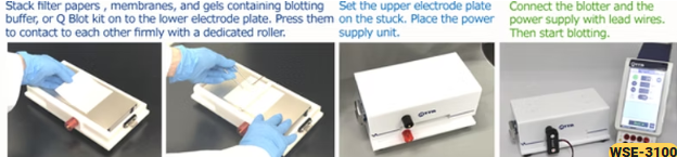
3- Blotting System

4- Gel Documentation Imaging System
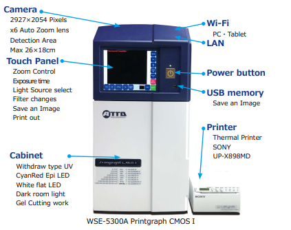
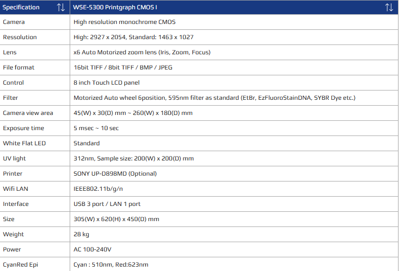
A-WSE-5300 Printgraph 6M
- UV Light Source (312 nm): Ideal for visualizing nucleic acid gels stained with Ethidium Bromide (EtBr), SYBR Green, SYBR Safe, and other fluorescent dyes.
- White Flat LED: Designed for imaging Coomassie Brilliant Blue (CBB) stained gels, silver-stained gels, and other colorimetric stains used in protein analysis.
- CyanRed Epi Illumination: A UV-free fluorescence detection system that offers significantly higher resolution than conventional blue LED systems, enabling clearer visualization of fluorescent signals without DNA damage risks.
- Direct Printout Capability: Compatible with high-quality digital graphic printers such as the SONY UP-D898MD for immediate hard copy generation.
- Advanced Image Saving: Store images directly on the main unit, export to USB memory, or remotely control and send images to tablets via WiFi connectivity
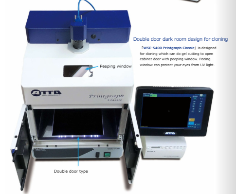
B-WSE-5400 Printgraph Classic
- UV Light Source: Ideal for capturing high-quality images of EtBr-stained gels, SYBR Green, and SYBR Safe stained gels commonly used in nucleic acid electrophoresis.
- White LED Plate Light Source (Optional): Designed for imaging Coomassie Brilliant Blue (CBB) stained gels, silver-stained gels, and other protein gel staining methods.
- Cyan LED Light Source (Optional): Enables imaging of multiple gel stains with enhanced sensitivity and versatility.
- Selectable Color and Monochrome Modes: Flexibility to capture images in color or monochrome depending on experimental requirements.
- Instant Image Printing: Supports immediate printing of captured gel images for quick documentation and analysis.
- Image Saving: Allows direct saving of captured images to USB memory for easy transfer and long-term storage.
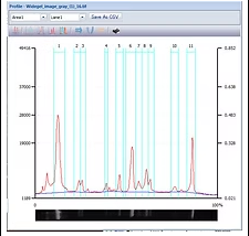
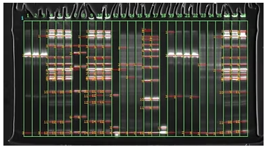
5- Image Analysis Software CS Analyzer 4
Purpose, application, and features
- Digitization of Electrophoresis Patterns: Efficiently capture and convert electrophoresis gel images into digital formats for detailed analysis.
- Comprehensive Analysis Results: Save and generate detailed reports based on electrophoresis data for accurate record keeping and study comparisons.
- Image Conversion and Saving: Seamlessly convert and save images in multiple formats for easy integration with laboratory databases and publications.
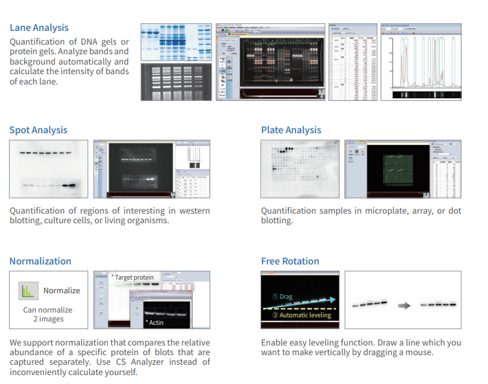
- LANE Analysis: Automated quantification and profiling for SDS-PAGE gels, agarose gels, and similar electrophoretic techniques.
- SPOT Analysis: Precise detection and quantification for Western blot spots and other immunodetection assays.
- PLATE Analysis: Specialized analysis for Dot blot and microplate-based assays.
- Housekeeping Protein Normalization: Reliable normalization to ensure accuracy in protein quantification.
- Molecular Weight Marker Calibration: Accurate sizing of proteins or nucleic acids using calibrated molecular weight markers.
- Scale Calibration: Precise image scale calibration for consistent measurement results.
- Image Mode Conversion: Flexible switching between color modes, including RGB merging and grayscale.
- Absorbance Conversion: Quantitative conversion of image data to absorbance values for enhanced analysis.
- Advanced Image Processing: Robust tools for image enhancement, background correction, and noise reduction.
- Data Management: Save, summarize, and export analysis data as comprehensive reports.
- Format Support: Compatible with common image formats including TIFF, BMP, JPEG, and proprietary CCD (ATTO format).
5- Reference LED Light Sources
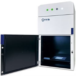
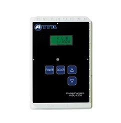
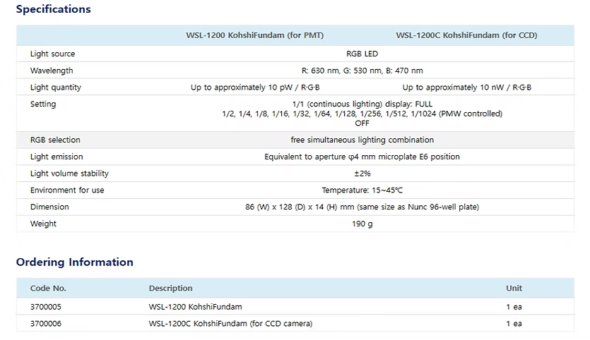
A-WSL-1200 KohshiFundam
Purpose, application, and features :
- Operational Qualification (OQ) Verification: Ensures imaging systems meet specified operational standards through rigorous testing.
- Performance Qualification (PQ) Verification: Confirms imaging systems consistently perform according to predetermined specifications under real-world conditions.
- Periodic Inspection: Facilitates regular checks of light measurement devices, including imaging systems, to maintain accuracy and reliability.
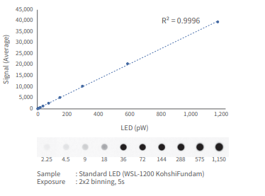
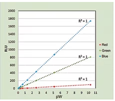
- Calibrated Reference Light Source: Calibrated against a standard reference light source, providing precise measurement of total light flux for reliable benchmarking.
- Stable RGB LED Emission: Utilizes a stable RGB LED light source adjustable from 10 nW to 10 fW, suitable for sensitive imaging and faint light measurement.
- Routine Equipment Checks: Ideal for regular verification and quality control of imaging systems and low-light measurement devices.
- PWM-Controlled Brightness: Features 11-step Pulse Width Modulation (PWM) control for precise and flexible light intensity settings.
- Linearity Testing Support: Supports accurate linearity assessment of optical measurement equipment to ensure consistent performance.
- Customizable RGB Settings: Allows independent on/off control and light quantity adjustment for each RGB color channel.
- Temperature Compensation: Incorporates heat stabilization via temperature compensation to maintain consistent light output despite ambient temperature fluctuations.
- Enhanced Reproducibility: Ensures high reproducibility in measurements due to stable light output under varying environmental conditions.
B-WSL-1230/1235/1240 KohshiUni
Purpose, application, features
- Sensitivity Testing of Imaging Devices:Accurate sensitivity verification of imaging sensors such as CCD and CMOS cameras across a wide light intensity range from nanowatts (nW) to microwatts (μW).
- Photomultiplier Tube (PMT) Sensitivity Checks:Precise sensitivity calibration for PMTs, effective in detecting extremely low light intensities from femtowatts (fW) to picowatts (pW).
- Detector Verification and Calibration:Comprehensive verification and calibration services for imaging and light detection equipment to ensure optimal performance and measurement accuracy.
Features
- Calibrated Reference Light Source: Light source calibrated by comparison with a standard reference, providing precise measurement of total light flux for dependable benchmarking.
- Stable LED Emission: Employs a highly stable LED light source adjustable to a total light intensity range of nW to μW for CCD/CMOS models, and fW to pW for PMT models.
- Routine Equipment Validation: Perfect for regular sensitivity checks and performance verification of imaging systems and low-light measurement instruments.
- PWM Light Intensity Control: Features 10-step Pulse Width Modulation (PWM) for accurate and repeatable adjustment of light intensity.
- Linearity Testing Support: Supports rigorous linearity testing of optical measurement devices to validate detector responses over varying intensities.
- Temperature Compensation: Incorporates advanced temperature compensation to stabilize light output, ensuring consistent results despite ambient temperature fluctuations.
- High Reproducibility: Guarantees excellent reproducibility in sensitivity tests due to stable light emission even under changing environmental conditions.