
Mineralocorticoid Receptor (MR; NR3C2) | IB00501
 | IB00501")
- SKU:
- IB00501-32 | IB00501 | IB00502
- Availability:
- In Stock
- Storage:
- -80°C
- Shipping:
- -80°C (International: -80°C)
 | IB00501")
Description
Mineralocorticoid Receptor (MR; NR3C2) | IB00501
This Mineralocorticoid Receptor (MR) assay kit is an all-inclusive MR reporter assay system that includes, in addition to MR Reporter Cells, two optimized media for use during cell culture and (optionally) in diluting the test samples, a reference agonist, Luciferase Detection Reagent, a cell culture-ready assay plate, and a detailed protocol.
MR Reporter Cells are prepared using INDIGO’s proprietary CryoMite™ process. This cryo-preservation method yields high cell viability post-thaw, and provides the convenience of immediately dispensing healthy, division-competent reporter cells into assay plates. There is no need for intermediate spin-and-wash steps, viability determinations, or cell titer adjustments.
INDIGO’s Mineralocorticoid Receptor assay kits feature a luciferase detection reagent specially formulated to provide stable light emission between 5 and 90+ minutes after initiating the luciferase reaction. Incorporating a 5-minute reaction-rest period ensures that light emission profiles attain maximal stability, thereby allowing assay plates to be processed in batch. By doing so, the signal output from all sample wells, from one plate to the next, may be directly compared within an experimental set.
Mineralocorticoid Receptor assay kits are offered in different assay formats to accommodate researchers’ needs: 3x 32, 1x 96, and 1x 384 assay formats for screening small numbers of test compounds, as well as custom bulk reagents for HTS applications.
Assay Kit & Platforms
Bulk assay reagents can be custom manufactured to accommodate any scale of HTS. Please inquire.
Assay Services
The primary application of INDIGO’s cell-based nuclear receptor assays are to quantitatively assess the bioactivity of a test compound as an agonist (activator) or antagonist (inhibition of an agonist response) of a given receptor. Service assays include a positive control reference compound and ‘vehicle’ control for every experiment. A formal study report and all data files are provided to the client upon completion of the study. To receive a quote for your proposed study, complete & submit the online “Request a Quote” form or contact an INDIGO Customer Service Representative to discuss your desired study parameters.
Background
The mineralocorticoid receptor (MR), also called aldosterone receptor, is a receptor with high affinity for mineralocorticoids. It belongs to the steroid hormone receptor family where the ligand diffuses into cells, interacts with the receptor, and results in a signal transduction affecting specific gene expression in the nucleus. The gene for the NR3C2 (located on chromosome 4q31.1-31.2) encodes for the 107 kDa MR protein.
MR is expressed in many tissues, such as the kidney, colon, heart, central nervous system (hippocampus), brown adipose tissue, and sweat glands. In epithelial tissues, its activation leads to the expression of proteins regulating ionic and water transports (mainly the epithelial sodium channel or ENaC, Na+/K+ pump, serum and glucocorticoid induced kinase or SGK1) resulting in the reabsorption of sodium, and as a consequence an increase in extracellular volume, increase in blood pressure, and an excretion of potassium to maintain a normal salt concentration in the body.
The receptor is activated by mineralocorticoids such as aldosterone and deoxycorticosterone as well as glucocorticoids, like cortisol and cortison. It also responds to some progestins. Spironolactone and eplerenone are mineralocorticoid receptor antagonists. Activation of the mineralocorticoid receptor, upon the binding of its ligand aldosterone, results in its translocation to the cell nucleus, homodimerization and binding to hormone response elements present in the promoter of some genes. This results in the complex recruitment of the transcriptional machinery and the transcription into mRNA of the DNA sequence of the activated genes.
INDIGO's Mineralocorticoid Receptor Reporter Assay Systems utilize proprietary mammalian cells engineered to express human NR3C2 protein, commonly referred to as MR.
The principle application of this assay product is in the screening of test samples to quantify functional activities, either agonist or antagonist, that they may exert against the human mineralocorticoid receptor.
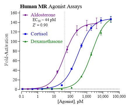
Agonist dose-response analyses of the Human MR. Analyses of MR Reporter Cells using Aldosterone (provided), Cortisol (Tocris), and Dexamethasone (Cayman Chemical). Concentrated stocks of agonists were prepared in DMSO, then serially diluted using CSM. Final assay concentrations for each agonist were: 4000, 1000, 250, 62.5, 15.6, 3.91, 0.977 and 0 pM. Luminescence was quantified and average relative light units (RLU) and corresponding standard deviation (SD), % coefficient of variation (%CV), and Fold-Activation values were determined for each treatment concentration (n = 4). Z’ values were calculated as described by Zhang, et al. (1999). Non-linear regression and EC50 analyses were performed using GraphPad Prism software. The low %CV, high S/B and exceptional Z' scores confirm the robust performance of this MR Assay and its suitability for HTS Applications.
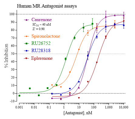
Validation of Antagonist-mode MR Assays. Human MR antagonist assay performance was validated using the reference antagonists Canrenone (Cayman Chemical), Spironolactone, RU26752, RU28318 and Eplerenone (all from Tocris). Assay setup and quantification of MR activity were performed following the protocol described in the assay Technical Manual. Human MR Reporter cells were co-treated with a fixed ~EC80 concentration of Aldosterone (140 pM) and varying concentrations of the respective antagonist compounds. Final assay concentrations of Canrenone, Spironolactone, RU28318 and Eplerenone ranged between 10 M and 150 pM. Treatment concentrations of RU26752 ranged from 80 nM to 10 pM. 'No antagonist' controls were included. Assay plates were incubated for 22 hrs, then processed to quantify MR activity for each treatment condition.
Related Products
Related Products

BV-MR-LBD Recombinant baculovirus expressing Mineralocorticoid receptor LBD | S27
-NR3C2 Plasmid")
pcDNA3.1(+)-NR3C2 Plasmid

pDONR223-NR3C2 Plasmid

pECMV-Nr3c2-m-FLAG Plasmid
")