
Rho Activator II | CN03
- SKU:
- CN03-A | CN03-B
- Availability:
- In Stock
Description
Rho Activator II | CN03
Background Information
The G-switch™ line of small G-protein tools has been developed with an emphasis on creating highly potent reagents that target endogenous Rho family proteins and pathways. In contrast to methods that rely on over-expression or knockdown of target proteins (e.g., DNA transfection of dominant negative or constitu-tively active Rho mutants, RNAi knockdown) , the G-switchTM reagents act rapidly on the endogenous target protein (in minutes to hours, depending on product), thereby optimizing the chance of generating a more physiologically relevant response. The G-switch™ product line includes reagents that directly and indirectly modulate Rho family signal transduction, thereby offering a wide range of mechanistic tools to study these critical cellular functions. See Cytoskeleton’s web site for the latest G-switch™ information.
The active site of CN03 is based on the catalytic domain of the bacterial cytotoxic necrotizing factor (CNF) toxins. The catalytic domain is covalently attached to a proprietary cell penetrating moiety. CN03 activates Rho GTPase isoforms by deamidating glutamine-63, which is located in the Switch II region of these GTPases (1). This modification converts glutamine-63 to gluta-mate, which blocks intrinsic and GAP-stimulated GTPase activity, resulting in constitutively active Rho (2). CN03 robustly increases the level of GTP-bound RhoA within 2-4 h after addition to the culture medium. Moreover, the targeted action of this activator makes it a far more attractive tool for the study of Rho GTPase signaling than classic indirect activators (e.g., LPA) that concomi-tantly activate other signaling pathways (e.g., Ras, PI3K and PLC).
Material
CN03 has been produced in a bacterial expression system. The recombinant protein has a mol wt of 118 kDa and contains six histidine residues at its amino terminus (His tag). It has been purified to >80% purity using immobilized metal affinity chromatog-raphy (Fig. 1). Supplied as an off-white lyophilized solid, each vial contains 20 µg of CN03 protein. The material has been shown to be active in a biological assay for RhoA activation (see below). A protein containing a single point mutation that inactivates the catalytic domain of CN03 showed zero activity in a biological assay for RhoA activation (data not shown).
Storage and Reconstitution
Shipped at ambient temperature. The lyophilized protein can be stored desiccated at 4°C for 6 months. The lyophilized pellets of CN03 may appear loose and lightly packed, but this does not affect the quality or performance of CN03. For reconstitution, briefly centrifuge to collect the product at the bottom of the tube and resuspend each vial in 200 µl of sterile water, place on ice for 10 minutes prior to mixing, and mix by gently pipetting up and down to yield a concentration of 0.1 µg/µl. Prior to use, CN03
should be further diluted with serum free growth medium to a concentration between 0.25 and 5 µg/ml (to be determined by user, also see Fig. 2 & Table 1). Typically, a 1 µg/ml concentra-tion produces robust activation of RhoA in multiple cell types within 3 h. Reconstituted activator can be snap frozen in liquid nitrogen and stored at –70°C for up to 6 months.
Biological Activity Assay
CN03 (1 ug/ml / equivalent to 10 µl/ml) was shown to induce a ten fold RhoA activation (Figs. 2 & 3) and stress fiber induction (Fig. 4) in Swiss 3T3 cells after a 2 h incubation at 37°C. Recommended conditions for Rho activation in several cell types are detailed in Table 1.
Activity Assay Method: Swiss 3T3 cell activation
1. Grow Swiss 3T3 cells at 37°C / 5% CO2 to 30% confluency in two 10 cm2 dishes containing 10 ml DMEM / 10% fetal bovine serum (FBS).
2. Serum starve cells by changing media to DMEM / 1% FBS for 24 h and then transferring to DMEM / 0% FBS for 24 h.
3. Briefly spin tube of CN03 to collect contents on bottom of tube.
4. Reconstitute CN03 with 200 µl of sterile water (0.1 µg/µl) - See Storage and Reconstitution section above.
5. Dilute CN03 to 1 µg/ml with warm DMEM.
6. Aspirate medium from both dishes of cultured cells and transfer CN03 containing medium onto one dish.
7. Transfer warm DMEM only to the second dish. This is the control and represents unstimulated cells.
8. Incubate for 2 h at 37°C and 5% CO2 . Assay Rho activity by G-LISA® analysis (Cat. # BK124; Figs 2 & 3) or cell morphology ( Cat. # PHDG1; Fig. 4)
Table 1. Suggested Conditions for Rho GTPase activation
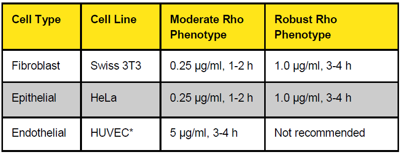
Legend: The indicated cells were subjected to RhoA, Rac1 and Cdc42 G-LISA® activation assays with CN03 in serum free medium. No Rac or Cdc42 activation was observed under these conditions. Note: A moderate Rho activation phenotype is characterized as an 80-150% increase in RhoA accompanied by a moderate increase in actin stress fibers. A robust phenotype is characterized by >400% increase in RhoA activation with cells exhibiting strong stress fiber morphology (see Fig.4). (*) Significantly weaker activity has been observed for CN03 in endothelial cells relative to other cell types.
Figure 1. Purity analysis of CN03 protein.
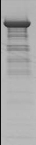
Legend: A 10 µg sample of CN03 protein was separated by electrophoresis in a 4-20% SDS -PAGE system and stained with Coomassie Blue. Densitometric analysis determined the protein to be >80% pure. Arrow shows CN03, faint bands under CN03 represent minor degredation products (small arrows).
Protein quantitation was performed using the Precision Red Protein Assay Reagent (Cat. # ADV02).
Figure 2. Dose-dependent activation of RhoA in Swiss 3T3 cells.
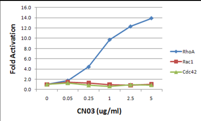
Legend: Swiss 3T3 fibroblasts were grown for 2 days in DMEM plus 10% FBS and serum starved for 24 h in media containing 1% FBS followed by 24 h in serum free media. Cells were treated with 0.05 to 5 μg/ml CN03 for 2 h and cell lysates were prepared using G-LISA® lysis buffer (Part# GL36) and were subjected to G-LISA® activation assays for RhoA (Cat.# BK124), Rac1 (Cat.# BK128) and Cdc42 (Cat.# BK127). Fold activation for each small G-protein was calculated from background-subtracted data.
Figure 3. Time-dependent activation of RhoA in Swiss 3T3 cells.
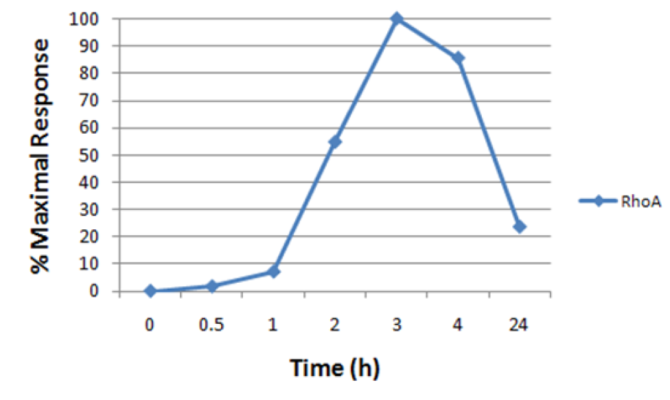
Legend: Swiss 3T3 fibroblasts were grown for 2 days in DMEM plus 10% FBS and serum starved for 24 h in media containing 1% FBS followed by 24 h in serum free media. Cells were treated with 1 μg/ml CN03 for the indicated times and cell lysates were prepared using G-LISA® lysis buffer (Part# GL36) and were sub-jected to G-LISA® activation assays for RhoA (Cat.# BK124). Data were normalized to the maximal activation level for each small G-protein to clearly show the time course of activation.
Figure 4. Actin cytoskeleton morphological changes induced by CN03 treatment of Swiss 3T3 cells.
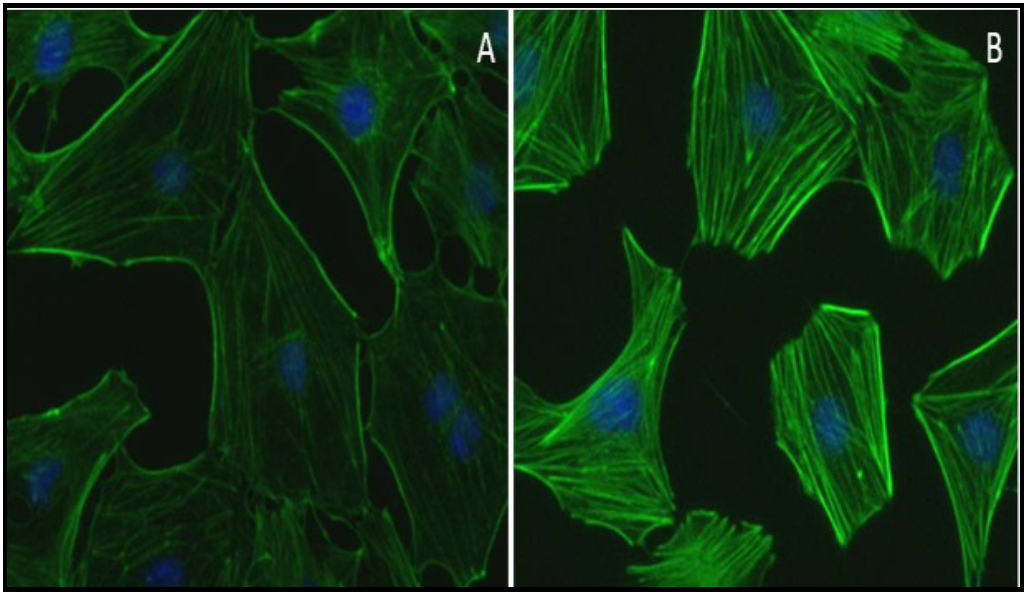
Legend: Swiss 3T3 fibroblasts were plated on glass coverslips, grown to 30% confluency in DMEM plus 10% FBS and serum starved for 24 h in media containing 1% FBS followed by 24 h in serum free media. Cells were treated with a buffer control (A) or 1 μg/ml CN03 for 2 h at 37°C/5% CO2 (B). Cells were then fixed, stained with Acti-stainTM 488 phalloidin (Cat.# PHDG1), and visualized by fluorescence microscopy. Images were taken at a magnification of 40x. The control cells (A) exhibited very few stress fibers, whereas treatment with CN03 (B) resulted in the development of abundant stress fibers. Under similar conditions, RhoA was activated ~10-fold as determined using the RhoA G-LISA® activation assay (Cat.# BK124) (See Fig. 2).
Product Uses
Rho pathway studies in cultured cells.
Study effects of altered Rho pathway signaling on other connected pathways.
Use as an active control when testing the effect of other substances on Rho pathway signaling.
Related Products
Related Products


Active-B12 EIA

ACTIVITY Aggrecanase Assay

RIPK1, Active | R07-10G
