Neuropeptide Y Antibody | 22940

(No reviews yet) Write a Review
SKU:
484-22940
Quantity/Volume:
100 µL
  • Neuropeptide Y Antibody | 22940
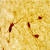
  • IHC image of neurons staining for neuropeptide Y in the rat cortex and striatum . The tissue was fixed with 4% formaldehyde in phosphate buffer, before being removed and prepared for vibratome sectioning. Floating sections were incubated at RT in 10% goat serum in PBS, before standard IHC procedure. Primary antibody was incubated at 1:5000 for 48 hours, goat anti-rabbit secondary was subsequently added for 1 hour after washing with PBS. Light microscopy staining was achieved with standard biotin-streptavidin/HRP procedure and DAB chromogen.
 
 The second image above entitled “C.” is NPY interneurons (RED) in mouse striatum, GREEN staining is GFP expressed by medium spiny neurons provided Alipi Naydenov, Neurobiology and Behavior, Stella Lab University of Washington (see review below).
  • High magnification of the globus pallidus with nickel preparation and low magnification IHC image of methionine encephalin staining in the globus pallidus and amygdala for the rat brain . The tissue was fixed with 4% formaldehyde in phosphate buffer, before being removed and prepared for vibratome sectioning. Floating sections were incubated at RT in 10% goat serum in PBS, before standard IHC procedure. Primary antibody was incubated at 1:2000 for 48 hours, goat anti-rabbit secondary was subsequently added for 1 hour after washing with PBS. Light microscopy staining was achieved with standard biotin-streptavidin/HRP procedure and DAB chromogen.
Select Currency //379.00

 Select your currency from the header

Description

Neuropeptide Y Antibody | 22940 | ImmunoStar

Neuropeptide Y (NPY) is a member of a regulatory peptide family and has a marked sequence homology with pancreatic polypeptide (PP) and peptide YY (PYY), which are other members of the family. NPY is widely expressed in the nervous system and has been shown to be differentially expressed in inhibitory interneurons in the hippocampus in degenerative disease, as a powerful vasoconstrictor in the periphery, and increased expression of NPY in the hypothalamus correlates with food intake. In the rat central nervous system, immunohistochemistry has found NPY-like cell bodies in the cortex, caudate-putamen, hypothalamus (arcuate nucleus), hippocampus, anterior westolfactory bulb, nucleus accumbens, amygdaloid complex and periaqueductal grey. NPY-like fibers and terminals are detected in high numbers in the bed nucleus of the stria terminalis, the peri- and paraventricular regions of the hypothalamus and thalamus and in discrete hypothalamic nuclei, particularly the suprachiasmatic nucleus. Westerns are not recommended for the Neuropeptide Y antibody since the protein is too small to detect in gel.

Host: Rabbit

State: Lyophilized Whole Serum

Reacts With: Alpaca (Llama), Bird, Bubalus Bubalis (Water Buffalo), Bullfrog, Cat, Ewe, Ferret, Fish, Frog, Frog (Xenopus Laevis), Ground Squirrel, Guinea Pig, Hamster, Human, Monkey, Mouse, Ovine (Sheep), Pig, Psetta Maxima (Flat Fish), Rat, Sheep, Snake, Tadpole, Toad, Turtle, Zebrafish

Availability: In Stock

Alternate Names: Pro-neuropeptide Y; Neuropeptide tyrosine; prepro-neuropeptide Y; PYY4, anti-NPY

Gene Symbol: NPY

View AllClose

0 Reviews

View AllClose