Mouse Anti OVA Monoclonal IgE Antibody E-C1, 1 mg, lyophilized
- SKU:
- 445-3006
- Size:
- 1 mg
- Shipping:
- Gel Packs
- Storage:
- -20 C
Description
Mouse Anti OVA Monoclonal IgE Antibody E-C1, 1 mg, lyophilized - Cat Number: 3006 From Chondrex.
Research Field: Allergy
Clonality: N/A
Cross-Reactivity:
Host Origin: N/A
Applications: N/A
Isotype: N/A
Detection Range: N/A
Sample Type: N/A
Concentration: N/A
Immunogen:
DESCRIPTION: Mouse anti-OVA IgE monoclonal antibody, clone E-C1, unconjugated
APPLICATION: Use for animal studies, ELISA, and Western Blotting
QUANTITY: 1 mg containing 1 mg mouse serum albumin as a stabilizer (no preservative)
FORM: Lyophilized powder
SOURCE: Mouse
IMMUNOGEN: Ovalbumin
CROSS-REACTIVITY: Cross-reacts to both native and aggregated OVA
PURITY: Purified by ion-exchange chromatography
SUBTYPE: Mouse IgE
USAGE: In vivo studies: reconstitute antibody with PBS containing 1 mg/ml MSA or with Chondrex, Inc.’s
IgE Dilution Buffer (Cat # 3009)
In vitro studies: reconstitute antibody with a buffer containing 2% heterologous serum, such as
normal goat serum, to prevent non-specific reactions.
STORAGE: -20⁰C
STABILITY: 1 year
NOTES: E-C1 can also be found in published literature under the clone name "OE-1". They are identical monoclonal antibodies.
REFERENCES: N. Mizutani, H. Goshima, T. Nabe, S. Yoshino, Complement C3a-induced IL-17 plays a critical role in an IgE-mediated late-phase asthmatic response and airway hyperresponsiveness via neutrophilic inflammation in mice. J Immunol 188, 5694-705 (2012).
N. Mizutani, H. Goshima, T. Nabe, S. Yoshino, Establishment and characterization of a murine model for allergic asthma using allergen-specific IgE monoclonal antibody to study pathological roles of IgE. Immunol Lett 141, 235-45 (2012).
N. Mizutani, C. Sae-Wong, S. Kangsanant, T. Nabe, S. Yoshino, Thymic stromal lymphopoietininduced interleukin-17A is involved in the development of IgE-mediated atopic dermatitis-like skin lesions in mice. Immunology 146, 568-81 (2015).
Y. Okamoto-Uchida, R. Nakamura, Y. Matsuzawa, M. Soma, H. Kawakami, et al., Different
Results of IgE Binding- and Crosslinking-Based Allergy Tests Caused by Allergen Immobilization. Biol Pharm Bull 39, 1662-1666 (2016).
C. Sae-Wong, N. Mizutani, S. Kangsanant, S. Yoshino, Topical skin treatment with Fab fragments of an allergen-specific IgG1 monoclonal antibody suppresses allergen-induced atopic dermatitis-like skin lesions in mice. Eur J Pharmacol 779, 131-7 (2016).
Chondrex, Inc. introduces a new protocol to induce nasal hypersensitivity in mice. Dr. Saika et al., published this model using Chondrex, Inc.’s anti-OVA IgE monoclonal antibody, E-C1, in C57BL/6 mice (1). Because many transgenic mouse strains are available, this model can be adapted to investigate the role of gene function in the development of allergic reactions. The evaluation methods are simple and do not require sacrificing mice. As a result, multiple evaluations can be conducted as well as an endpoint study, allowing complete evaluation of the entire study period. The following information is a summary of the protocol and results. Please contact Chondrex, Inc if you are interested in the anti-OVA IgE monoclonal antibody (Cat # 3006).
MATERIALS AND METHODS
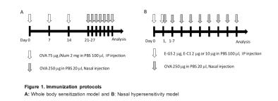
Experimental Animals C57BL/6JJcl male mice were acclimated for a week in Specific Pathogen Free conditions at room temperature (23 ± 3°C), humidity 30 to 90%, lighting time 7:00 to 21:00, and allowed to ingest solid food and water ad libitum. Whole-body Sensitization Model The mouse allergic rhinitis model was induced using egg white albumin (OVA) as the allergen. The mice received 2 mg of Aluminum adjuvant containing 75 μg of OVA in PBS
intraperitoneally on days 0, 7, and 14. Then the mice received 20 μl of PBS containing 250 μg of OVA in both nasal passages every day from day 21 to 27 (OVA/OVA group). 24 hours after the last sensitization, serum and tissue samples were collected from mice (Figure 1A). Negative control mice received only PBS administered intraperitoneally/intranasally (PBS/PBS group).
Nasal Hypersensitivity Model
Mouse anti-OVA-specific IgE antibodies (Clones E-C1 and E-G5, Cat # 3006 and 3007, respectively, Chondrex, Inc.) were used. Mice received E-C1 (2 μg or 10 μg) or E-G5 (2 μg) in PBS,i ntravenously, on days 0 and 1. Then the mice received 20 μl of PBS containing 250 μg of OVA in both nasal passages every day from day 1 to 7 (OVA/OVA group) (Figure 1B). Negative control mice received only PBS administered intravenously/intranasally (PBS/PBS group). Evaluating Symptoms After the final intranasal sensitization, the number of times that nasal scratching and sneezing occurred was measured for 10 minutes.
Measuring Serum OVA-specific IgE
Serum was collected on day 28 in the whole-body sensitization models and day 8 in the nasal hypersensitivity models. Mouse serum anti-OVA IgE levels were analyzed with an ELISA kit.
Histological Evaluation
The heads were fixed in 4% paraformaldehyde, demineralized with 0.5 M ethylenediamine tetraacetic acid for 1 week. The samples were then embedded in paraffin and sliced 6 μm thick to make slide samples.
The slides were stained with Periodic acid Schiff (PAS), and the number of goblet cells in a 100 μm wide section of basement membrane were counted in three arbitrarily selected regions of the nasal septum mucosa. In addition, the thickness of the nasal mucosa was measured in three randomly selected regions.
RESULTS
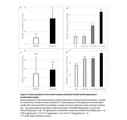
In the whole-body sensitization model, the number of sneezing incidents was significantly higher in the OVA/OVA group (21.0 ± 7.1 times) compared to the PBS/PBS group (4.8 ± 1.5 times) (Figure 2A). The number of nasal scratching incidents tended to be higher in the OVA/OVA group (37.8 ± 12.2 times) than in the PBS/PBS group (28.8 ± 37.8 times) (Figure 2C). In addition, the mice who received OVA sensitization and PBS nasal administration showed no nasal allergy symptoms (OVA/PBS data not shown). In the nasal hypersensitivity model, the number of scratching and sneezing incidents in the mice who received 10 μg E-C1 was higher than the three other groups who received 2 μg E-C1, 2 μg E-G5, or PBS (Figure 2B and 2D).
