
Mouse Anti OVA Monoclonal IgE Antibody E-G5, 1 mg, lyophilized

- SKU:
- 445-3007
- Size:
- 1 mg
- Shipping:
- Gel Packs
- Storage:
- -20 C

Description
Mouse Anti OVA Monoclonal IgE Antibody E-G5, 1 mg, lyophilized - Cat Number: 3007 From Chondrex.
Research Field: Allergy
Clonality: N/A
Cross-Reactivity:
Host Origin: N/A
Applications: N/A
Isotype: N/A
Detection Range: N/A
Sample Type: N/A
Concentration: N/A
Immunogen:
DESCRIPTION: Mouse anti-OVA IgE monoclonal antibody, clone E-G5, unconjugated
APPLICATION: Use for animal studies, ELISA, and Western Blotting
QUANTITY: 1 mg containing 1 mg mouse serum albumin as a stabilizer (no preservative)
FORM: Lyophilized powder
SOURCE: Mouse
IMMUNOGEN: Ovalbumin
CROSS-REACTIVITY: Reacts to native OVA
PURITY: Purified by ion-exchange chromatography
SUBTYPE: Mouse IgE
USAGE: In vivo studies: reconstitute antibody with PBS containing 1 mg/ml MSA or with Chondrex, Inc.’s
IgE Dilution Buffer (Cat # 3009)
In vitro studies: reconstitute antibody with a buffer containing 2% heterologous serum, such as
normal goat serum, to prevent non-specific reactions.
STORAGE: -20⁰C
STABILITY: 1 year
NOTES: N/A
REFERENCES: N. Mizutani, H. Goshima, T. Nabe, S. Yoshino, Complement C3a-induced IL-17 plays a critical role in an IgE-mediated late-phase asthmatic response and airway hyperresponsiveness via neutrophilic inflammation in mice. J Immunol 188, 5694-705 (2012).
N. Mizutani, H. Goshima, T. Nabe, S. Yoshino, Establishment and characterization of a murine model for allergic asthma using allergen-specific IgE monoclonal antibody to study pathological
roles of IgE. Immunol Lett 141, 235-45 (2012).
N. Mizutani, C. Sae-Wong, S. Kangsanant, T. Nabe, S. Yoshino, Thymic stromal lymphopoietininduced interleukin-17A is involved in the development of IgE-mediated atopic dermatitis-like
skin lesions in mice. Immunology 146, 568-81 (2015).
Y. Okamoto-Uchida, R. Nakamura, Y. Matsuzawa, M. Soma, H. Kawakami, et al., Different
Results of IgE Binding- and Crosslinking-Based Allergy Tests Caused by Allergen
Immobilization. Biol Pharm Bull 39, 1662-1666 (2016).
C. Sae-Wong, N. Mizutani, S. Kangsanant, S. Yoshino, Topical skin treatment with Fab fragments of an allergen-specific IgG1 monoclonal antibody suppresses allergen-induced atopic dermatitis-like skin lesions in mice. Eur J Pharmacol 779, 131-7 (2016).
INTRODUCTION
Type I hypersensitivity, which is characterized by an allergic reaction immediately following contact with innocuous antigens, is a typical clinical feature of allergic diseases such as asthma, eczema, hay fever, or urticaria. This hypersensitivity mediated by the IgE antibodies is called “atopic reagin” and the clinical features of type I hypersensitivity are described as “atopy”. The high affinity receptor for IgE (FcεRI) expressed on mast cells and basophils is another critical component in allergic responses. IgE bound to FcεRI drastically up-regulates FcεRI expression on mast cells through stabilization and accumulation of FcεRI and contributes in further enhancing hypersensitivity responses to allergens (1). Specific allergens bound to IgE on cell surfaces will cross-link two FcεRIs and this linkage leads to the stimulation and degranulation of mast cells (2-3). This is associated with the release of a variety of proinflammatory mediators and cytokines such as histamine, proteolytic enzymes, heparin, and chemotactic factors, which cause the symptoms associated with type I hypersensitivity.
Mice are widely used experimental animals for studying pathogenesis and evaluating therapeutics in allergic disease models as a variety of inbred strains, transgenic mice, and even gene knockout mice are available (2-6). In order to induce allergic diseases in animals, immunize animals with an antigen adsorbed on aluminum hydroxide gel or expose animals to an aerosolized antigen repeatedly (7-9). In addition, IgE antibodies against specific antigens can sensitize animals by passive transfer in vivo as well as sensitize mast cells in vitro (8-10). For example, passive transfer of airway hyper-responsiveness by ovalbumin (OVA)-specific monoclonal IgE and IgG antibodies was reported by Oshiba et al., although eosinophilic inflammatory response in the airway is modest despite high levels of serum IgE (8).
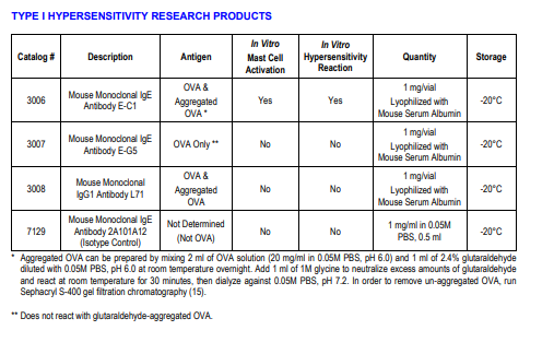
LIST OF IgE AND IgG MONOCLONAL ANTIBODIES AGAINST OVA
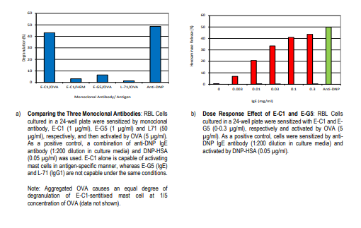
Chondrex, Inc. provides two IgE monoclonal antibodies specific to OVA, clone E-C1 and clone E-G6, in addition to one IgG1 monoclonal antibody against OVA, clone L71 (see Type I Hypersensitivity Research Products) (13-15). In general, the cross-linkage of IgE molecules bound to the receptor on mast cells by a polyvalent allergen is required to trigger degranulation of mast cells. Alternatively, IgG antibodies specific to the allergen can trigger the activation of mast cells by cross-linking two allergens bound by IgE antibodies on the mast cell surfaces (10). E-C1 alone is capable of inducing degranulation of mast cells in vitro (Figure 1), and severe hypersensitivity in vivo (Figure 2). Therefore, it is assumed that E-C1 might recognize repetitive epitopes of OVA. Furthermore, it has been reported that aggregated OVA, which carries multiple epitopes, increases the formation of cross-linked IgE on mast cell surfaces (16). On the other hand, E-G5 is not capable of inducing these hypersensitivity reactions by itself either in vitro or in vivo (Figures 1 and 2) and thus can be used as a control. Alternatively, Chondrex, Inc also provide a mouse IgE monoclonal antibody, isotype control, clone 2A101A12 as a negative control antibody.
The IgG1 monoclonal antibody, L71, can be used for a variety of research purposes such as studying the interference between allergen specific IgG and IgE antibodies as the pathogenic role of IgG in allergic diseases remains uncertain (11). For example, 1) IgG antibodies might compete with IgE antibodies and block the immediate hypersensitivity reactions, 2) IgG antibodies might stimulate IgE-mediated hypersensitivity reactions by cross-linking allergens bound to IgE on the surfaces of mast cells, or 3) IgG antibody-antigen immune complexes might stimulate IgE mediated hypersensitivity reactions by forming multivalent antigens which might bind to IgE on the surfaces of mast cells.
EXPERIMENTAL PROTOCOLS
A. Handling the IgE Antibody Solution
IgE antibody is biologically active at very low levels, so extra caution is required for handling and diluting IgE antibodies. In general, several 10 µg of protein will be adsorbed to 1 square cm of glass or plastic surfaces by non-specific hydrophobic binding. Therefore, if 1 ml of a 20 µg/ml IgE solution without blocking agents (such as PBS) is kept in a microcentrifuge tube, almost all the IgE antibodies will be adsorbed to the tube and no detectable IgE antibodies will remain in solution. Do not dilute IgE solutions to less than 100 µg/ml with PBS as it does not contain blocking agents. Add a suitable blocking agent to PBS, such as mouse serum albumin, bovine serum albumin (BSA), or other suitable proteins. IgE Dilution Buffer (Cat # 3009) is preferred for diluting IgE for both in vitro and in vivo experiments.
B. Assay for Degranulating Mast Cells In Vitro
1. Culture mast cells such as rat basophilic leukemia cells (RBL-2H3) until the stationary phase. Re-suspend the cells at 2.5 x 10⁵ cells/ml in Eagle’s minimum essential medium containing 5% fetal calf serum (FCS) and transfer cells into 96-well (250 µl: 6.25 x 10⁴ cell/well) or 24-well (1 ml: 2.5 x 10⁵ cells/well) flat bottom cell culture plates, and continue to culture at 37°C overnight.
2. Add IgE monoclonal antibodies diluted with PBS containing blocking agents or culture medium containing FCS to the wells (final concentration: 0-10 µg/ml of medium) and incubate for 1 hour.
3. NOTE: Instead of adding IgE into culture media after overnight cell culture, IgE can be added to culture media at the start of cell culture. In this case, FcεRI expression could be significantly upregulated with the overnight culture. For example, it has been reported that Fc Wash IgE-sensitized cells 3 times with Tyrode’s buffer (150 mmol/l NaCl, 2.5 mmol/l KCl, 12 mmol/l NaHCO3, 2 mmol/lMgCl2, 2 mmol/l aCl2, 1 mg/ml BSA, 1 mg/ml dextrose, pH 7.4).
5. Add 200 µl (in 96-well plate) or 400 µl (in 24-well plate) of OVA solution diluted with Tyrode’s buffer (the final concentration of antigen: 0-5 µg/ml) and culture for 1 hour.
6. Add 200 µl (in 96-well plate) or 400 µl (in 24-well plate) of a negative control antigen such as hen egg lysozyme (HEL) or BSA (5 µg/ml) dissolved in Tyrode’s buffer into the negative control wells to determine spontaneous release of β-hexosaminase.
7. Add 200 µl (in 96-well plate) or 400 µl (in 24-well plate) of 1% Triton X-100 solution dissolved in Tyrode’s solution into 100% control wells to determine the total release of β-hexosaminase.
8. Assay β-hexosaminase activity in the culture supernatant using p-nitro-phenyl-N-acetyl b-D-glucosaminide as a substrate (11). Figure 1 - Degranulation of Mast Cells by Monoclonal IgE Antibody.
Dilute IgE antibody solution (1 mg/ml) to 100 µg/ml with PBS containing a blocking agent such as mouse serum albumin. Inject 100 µl of the diluted IgE monoclonal antibody solution intravenously into mice (10 µg/mouse). Inject 25 µl of OVA (2 mg/ml in PBS) intradermally at the foot pad (50 µg/foot pad) after 24 hours. Determine the swelling of the footpad by measuring the footpad thickness using a dial thickness gauge every 1-2 hours for 24 hours.
Related Products
Related Products

Mouse Anti OVA Monoclonal IgE Antibody E-C1, 1 mg, lyophilized

Mouse Anti OVA Monoclonal IgG1 Antibody L71, 1 mg, lyophilized

Mouse Anti-Ovalbumin IgE Monoclonal Antibody, Clone E-G5

