
Hydroxyproline Assay Kit

- SKU:
- 445-6017
- Size:
- 1 kit
- Shipping:
- Gel Packs
- Storage:
- -20 C

Description
Hydroxyproline Assay Kit - Cat Number: 6017 From Chondrex.
Research Field: ECM
Clonality: N/A
Cross-Reactivity:
Host Origin: N/A
Applications: N/A
Isotype: N/A
Detection Range: 400 µg/ml-6.3 µg/ml
Sample Type: Tissue Homogenate, Cells, Collagen Solution
Concentration: N/A
Immunogen:
PRODUCT SPECIFICATIONS
DESCRIPTION: Assay kit to quantify total collagen content (regardless of type or species)
FORMAT: 96-well ELISA Plate with removeable strips
ASSAY TYPE: Colorimetric assay
ASSAY TIME: 1 hour
STANDARD RANGE: 400 µg/ml to 6.3 µg/ml
NUMBER OF SAMPLES: Up to 40 (duplicate) colorless samples/plate and up to 20 (duplicate) colored samples/plate
SAMPLE TYPES: Tissue homogenate and cell culture media
RECOMMENDED SAMPLE DILUTIONS: Not required
CHROMOGEN: N/A (read at 530-560 nm)
STORAGE: -20°C
VALIDATION DATA: Intra-assay (3.4-6.3%)/Inter-assay (5.1-7.6%)/Spiking Test (91-95%)
NOTES: Select samples may require a 24-hour hydrolysis step
INTRODUCTION
Collagen is the major structural protein of the extracellular matrix in many tissues. Hydroxyproline, a major component of collagen, makes
up about 13.5% of its amino acid composition. Due to its highly restricted distribution in collagen, the hydroxyproline content accurately
reflects the amount of collagen. Therefore, quantitating hydroxyproline has been utilized for evaluating tissue fibrosis or collagen deposition
(1 - 3). However, conventional hydroxyproline assays are not useful because they require cumbersome procedures and special tools.
Chondrex, Inc. offers a hydroxyproline assay kit (Cat # 6017) which employs an improved assay system that can be operated with ease and
precision using 96-well plates. This kit can quantify the total collagen content in any tissue specimen or tissue homogenate, regardless of
collagen type or species. Chondrex, Inc. also provides additional collagen detection kits for different purposes. Firstly, the Sirius Red Total
Collagen Detection Kit (Cat # 9062 or 9062P) can be used to quantify solubilized collagen in samples. Secondly, the collagen detection
ELISA kits specific to type I or II collagen of various species are used for distinguishing collagen types and species in samples. Lastly, the
Sirius Red/Fast Green Collagen Staining Kit (Cat # 9046) is a semi-quantitative assay to determine the total collagen content in tissue
sections. For more information, please visit www.chondrex.com or contact support@chondrex.com.
KIT COMPONENTS

Not included: concentrated HCl (10N) and glass screw-thread vials (1-2 ml) with Teflon caps (Example: National Scientific B7999-1)
ASSAY OUTLINE
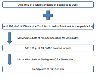
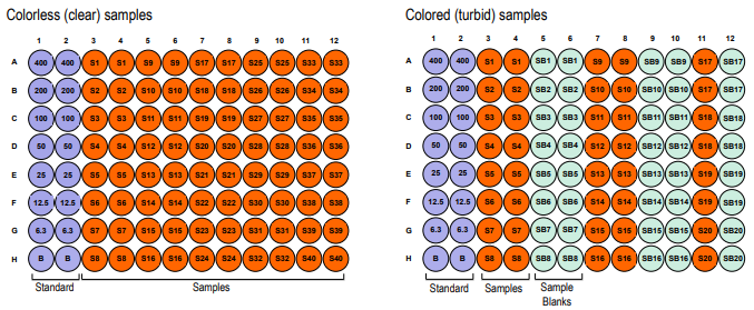
PLATE LAYOUT
NOTES BEFORE USING ASSAY
NOTE 1: It is recommended that the standard and samples be run in duplicate.
NOTE 2: For partial reagent use, please see the assay protocol’s corresponding step for the appropriate dilution ratio. For example, if the
protocol dilutes 50 µl of a stock solution in 10 ml of buffer for 12 strips, then for 6 strips, dilute 25 µl of the stock solution in 5 ml of buffer.
Partially used stock reagents may be kept in their original vials and stored at -20⁰C for use in a future assay.
NOTE 3: If there are precipitates in the bottles/vials, it is necessary to warm up the bottles/vials in warm water until they completely dissolve.
SAMPLE HYDROLYSIS
1. Weigh 10 mg of a tissue sample in a glass screw-thread vial.
2. Add 100 µl of distilled water.
3. Mash the tissue sample with a small spatula.
NOTE: 100 µl of a sample homogenate can be used. Skip steps 1-3 and directly add 100 µl of the sample homogenate to the vial.
4. Add 100 µl of concentrated HCl (10N) and tightly screw on the teflon cap.
5. Incubate at 120°C for 24 hours. Mix the sample periodically during incubation.
NOTE: A heat block or dry bath can be used for incubation.
6. Cool down. Do not open the cap before cooling down.
7. Use the supernatant for the assay.
NOTE: Black residue is occasionally produced from tissue samples in the hydrolysis process. However, sample hydrolyzation should
be complete by the end of the incubation period. If hydrolyzed black residue is still present in the sample, transfer to a microcentrifuge
tube and spin at 10,000 rpm for 3 minutes. Collect supernatant.
1 Review
-
Reliable and accurate hydroxyproline measurement
This kit offers excellent sensitivity and reproducibility, making it ideal for collagen-related research and fibrosis studies
Related Products
Related Products

Creatinine Assay Kit

Hemoglobin Assay Kit

DNA Assay Kit

Glycosaminoglycans Assay Kit
