
Type IX Collagen Staining Kit, Multi-Species

- SKU:
- 445-9012
- Size:
- 1 kit
- Shipping:
- Gel Packs
- Storage:
- -20 C

Description
Type IX Collagen Staining Kit, Multi-Species - Cat Number: 9012 From Chondrex.
Research Field: ECM
Clonality: N/A
Cross-Reactivity:
Host Origin: N/A
Applications: N/A
Isotype: N/A
Detection Range: N/A
Sample Type: Tissue Slides, Cultured Cells
Concentration: N/A
Immunogen:
PRODUCT SPECIFICATIONS
DESCRIPTION: Stain kits to detect Type II (Cat # 9001), Type IX (Cat # 9012), and Human Type I (Cat #9044) collagen
FORMAT: Glass slides
ASSAY TYPE: Immunostaining
ASSAY TIME: 3.5 hours
STANDARD RANGE: N/A
NUMBER OF SAMPLES: Varies
SAMPLE TYPES: Cultured cells and tissue sections
RECOMMENDED SAMPLE DILUTIONS: N/A
CHROMOGEN: Chondrex, Inc. recommends DAB
STORAGE: -20°C for 12 months
VALIDATION DATA: N/A
NOTES: Samples need to be prepared on slides before running assay
OVERVIEW
More than 26 distinct types of collagen have been identified in mammalian tissues. The collagen molecule is unique and composed of three alpha chains. The amino acid sequence is also unique and consists of repeated sequences characterized by Glycine-Proline-Hydroxyproline to form a stable, rigid triple helical structure. This structure is highly resistant to proteolytic degradation, heat, various chemicals, and physical stress. Tissue contribution of individual types of collagen is also unique. For example, type I collagen is most abundant in various connective tissues such as skin and bone. However, type I collagen does not exist in articular hyaline cartilage and the vitreous body of the eye. Instead, these tissues are composed of type II (80-85%), type IX (3-5%), and type XI collagen (5-10%). As the different types of collagen share similar amino acid sequences and three-dimensional structure, it is difficult and impossible to identify the collagen types in tissues by ordinary
chemical or histological means. To identify unique collagen types in connective tissues such as hyaline cartilage and other tissues, Chondrex, Inc. provides three types of collagen staining kits using collagen type specific monoclonal antibodies: 1) specific to many species of type II collagen, 2) specific to many species of type IX collagen and 3) specific to human type I collagen.
Type II Collagen Staining: A cocktail of biotinylated mouse monoclonal antibodies, Clone A2-10 (IgG2a), F10-21 (IgG2a), D8-6 (IgG2a), and D1-2G (IgG2b), recognizes the conserved epitopes of various species of type II collagen such as human, monkey, porcine, bovine, rat, mouse, rabbit, dog, equine, and chick and cross-reacts to all these species of type II collagen by more than 95%. These clones are highly specific to native type II collagen and poorly react to denatured collagen.
NOTE: As the α3(IX) chain of type XI collagen is identical to the α1(II) chain of type II collagen, these monoclonal antibodies may cross-react to type XI collagen, but not type I, type III, and type IX collagen. For immunoblotting, a 10X higher concentration of monoclonal antibodies will be required due to low reactivity with denatured collagen.
Type IX Collagen Staining: A cocktail of biotinylated mouse monoclonal antibodies, Clone D1-9 (IgG1) and B3-1 (IgG2b), recognizes the conserved epitopes on the low molecular weight fragment of various species of type IX collagen and equally cross-reacts to human, bovine, rat, and mouse type IX collagen, but only cross-reacts by 50% with chick type IX collagen in ELISA. These clones react with both native and denatured type IX collagen and can be used for both tissue staining and immunoblotting.
NOTE: Neither of these monoclonal antibodies reacts to type I, type II, or type XI collagen.
Human Type I Collagen Staining: A biotinylated rat monoclonal antibody, Clone 42R (IgM), recognizes a specific epitope on human type I collagen and does not react to other species of type I collagen such as porcine, bovine, rat, mouse, or chick.
NOTE: This clone is highly specific to native human type I collagen and poorly reacts to denatured collagen.
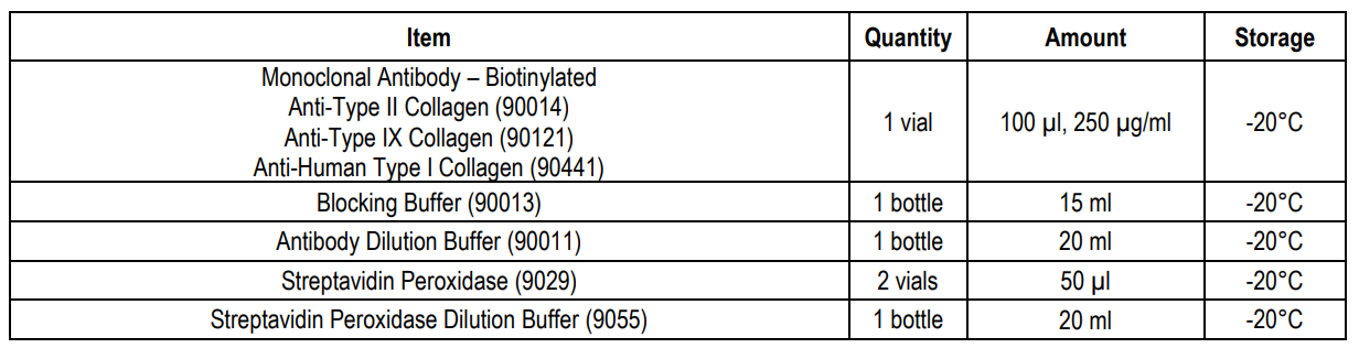
KIT COMPONENTS
REAGENTS AND SUPPLIES NOT PROVIDED
1. Glass coverslips or slides
2. Fixative (2.5% glutaraldehyde, 1% formalin, methanol (-20°C), or cold acetone)
3. If preparing formalin-fixed, paraffin-embedded tissue sections, xylene and ethanol is required.
4. 2% bovine testicular hyaluronidase (H3884 from Sigma-Aldrich is an example of one source of hyaluronidase.)
5. 0.1-1% hydrogen peroxide in methanol
6. Phosphate buffered saline (PBS)
7. Insoluble chromogen (such as DAB)
8. Mounting media
PREPARING SLIDES
A. Cultured Cells
1. Culture cells on sterile glass cover slips or slides.
2. Wash 3 times with PBS.
3. Fix cells by incubating with 2.5% glutaraldehyde in PBS for 10 minutes at 4°C.
4. NOTE: 1% formalin, methanol (-20°C), and cold acetone can be used for fixing samples. Please choose an appropriate method depending on samples.
5. Wash 3 times with PBS.
B. Frozen Tissue Sections
1. Prepare frozen tissues.
2. Prepare tissue sections (4-10 µm thick) and adhere sections onto egg white coated or Plus slides at room temperature.
3. Fix sections with 2.5% glutaraldehyde in PBS for 10 minutes at 4°C.
4. NOTE: 1% formalin, methanol (-20°C), and cold acetone can be used for fixing samples. Please choose an appropriate method depending on samples.
5. Wash 3 times with PBS.
C. Formalin-Fixed, Paraffin-Embedded Tissue Sections
1. Fix tissues in 1% formalin and embed in paraffin blocks.
2. Prepare tissue sections (4-10 µm thick) onto slides.
3. De-paraffinize 3 times with xylene for 5 minutes each.
4. Hydrate sections stepwise using ethanol solutions and PBS.
a. Wash with 100% ethanol for 5 minutes.
b. Wash with 95% ethanol for 5 minutes.
c. Wash with 75% ethanol for 1 minute.
5. Wash 3 times with PBS.
IMMUNOSTAINING
1. Treat sections with 2% bovine testicular hyaluronidase dissolved in PBS, pH 7.4 for 30 minutes at room temperature.
NOTE: Hyaluronidase removes glycosaminoglycan chains which may mask some tissue antigens, and thus facilitate binding of detection antibodies against collagens.
2. Wash 3 times with PBS.
3. Inactivate endogenous peroxidase by incubating with 0.1-1% hydrogen peroxide dissolved in methanol for 10 minutes.
4. Wash 3 times with PBS.
5. Incubate sections with Blocking Buffer for 30 minutes at room temperature.
6. Wash 3 times with PBS.
7. Dilute the monoclonal antibodies to type I, type II, or type IX collagen 1:250-1:2500 with Antibody Dilution Buffer.
8. Incubate the sections with diluted monoclonal antibody solution for 1 hour at room temperature.
9. Wash 3 times with PBS and gently shake off excess.
10. Dilute one vial of Streptavidin Peroxidase in 10 ml of Streptavidin Peroxidase Dilution Buffer.
11. Incubate the sections with diluted streptavidin peroxidase solution for 1 hour at room temperature.
12. Wash 3 times with PBS and gently shake off excess.
13. Develop the sections with an insoluble chromogen such as DAB (brown color).
14. Set slides with an immunohistochemical compatible mounting media and apply coverslips
Related Products
Related Products

Type II Collagen Staining kit, Multi-Species

Type II Collagen Detection Kit, Multi-Species

Human Type I Collagen Staining Kit
, polyclonal")
rb-Anti-r-Collagen Type IX (IgG), polyclonal
