Tubulin Polymerization Assay Using >99% Pure Tubulin, Fluorescence Based | BK011P
- SKU:
- BK011P
- Availability:
- In Stock
- Size:
- 96 assays
Description
Product Uses Include
- Cost effective high throughput screen for anti-cancer compounds.
- Basic research to measure compound IC50s and specificity for tubulin.
- Screening proteins for effects on tubulin polymerization activity.
- Teaching aid for undergraduate/graduate class in pharmacology.
Introduction
This assay is an economical one step procedure for determining the effects of drugs or proteins on tubulin polymerization. It is an adaptation of an assay originally described by Bonne, D. et al. (1). Polymerization is followed by fluorescence enhancement due to the incorporation of a fluorescent reporter into microtubules as polymerization occurs. The standard assay uses neuronal tubulin (Cat. # T240), which generates a polymerization curve representing the three phases of microtubule formation; namely nucleation, growth and steady state equilibrium. Other tubulins, such as HeLa cell tubulin (Cat. # H001) can also be used in this assay. The low volume per assay of 50 µl and the low tubulin concentrations, 2 mg/ml final concentration (100 µg per assay), makes this an ideal choice for studying the more expensive cancer cell tubulin reagents and for high throughput applications.
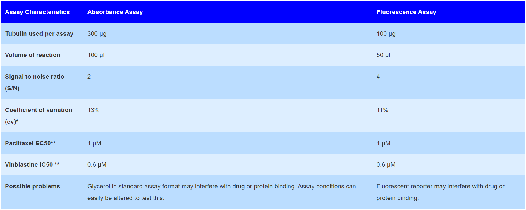
The classic tubulin polymerization assay uses absorbance readings at 340 nm to follow microtubule formation. It is based upon the fact that light is scattered by microtubules to an extent that is proportional to the concentration of microtubule polymer. This assay is offered by Cytoskeleton, Inc. (Cat. # BK006P). The fluorescence based assay has been compared to the absorbance based format and the comparisons are given in Table 1 below. For help in selecting the best assay format for your needs, contact info@gentaur.com
Table 1. Comparison of Fluorescence versus Absorbance Based Polymerization Assays

| *: Duplicate samples **: Under standard assay conditions. Conditions can be optimized for polymerization enhancers or inhibitors. |
Kit contents
The kit contains sufficient material for 96 assays in 50 µl format. The following components are included:
- >99% pure tubulin (Cat. # T240)
- General tubulin buffer with fluorescent reporter
- Microtubule glycerol buffer (Cat. # BST05)
- GTP solution (Cat. # BST06)
- Paclitaxel positive control (Cat. # TXD01)
- Half area 96-well plate. Black, flat bottom
- Manual with detailed protocols and extensive troubleshooting guide
Equipment needed
- Tempererature controlled 96-well plate fluorimeter equipped with filters for exitation at 340-360 nm and emission at 420-450 nm
Example results
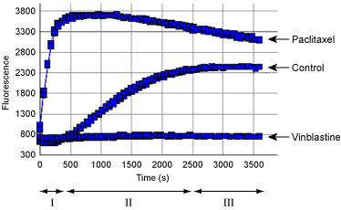
Compounds or proteins that interact with tubulin will often alter one or more of the characteristic phases of polymerization. For example, Figure 1 shows the effect of adding the anti-mitotic drug paclitaxel to the standard polymerization reaction. A 3 µM concentration of paclitaxel eliminates the nucleation phase and enhances the Vmax of the growth phase. Thus, one application of this assay is the identification of novel anti-mitotics. Figure 1 also shows the effect of adding the microtubule destabilizing drug, vinblastine. At 3 µM final concentration, vinblastine causes a drastic decrease in Vmax and reduction in final polymer mass.

Figure 1. Tubulin polymerization using the fluorescence based tubulin polymerization assay (BK011P). Tubulin was incubated alone (Control), with Paclitaxel or Vinblastine. Each condition was tested in duplicate. Polymerization was measured by excitation at 360 nm and emission at 420 nm. The three Phases of tubulin polymerization are marked for the control polymerization curve; I: nucleation, II: growth, III: steady state equillibrium.


