
Tubulin Protein (Fluorescent HiLyte 647): Porcine Brain | TL670M
: Porcine Brain | TL670M")
- SKU:
- TL670M-A | TL670M-B
- Availability:
- In Stock
: Porcine Brain | TL670M")
: Porcine Brain | TL670M")
: Porcine Brain | TL670M")
: Porcine Brain | TL670M")
Description
Tubulin Protein (Fluorescent HiLyte 647): Porcine Brain | TL670M
Product Uses Include
- Laser based applications
- Monitoring microtubule dynamcs in living cells
- Speckle microscopy
- Formation of fluorescent microtubules
- Microscopy studies of MAP and microtubule associated motor activities
- Nanotechnology
Material
Porcine brain tubulin (>99% pure, see Cat. # T240) has been modified to contain covalently linked HiLyte Fluor™ 647 (HiLyte Fluor is a trademark of Anaspec Inc, CA) at random surface lysines. An activated ester of HiLyte Fluor™ 647 was used to label the protein. Labeling stoichiometry was determined by spectroscopic measurement of protein and dye concentrations (dye extinction coefficient when protein bound is 250,000M-1cm-1). Final labeling stoichiometry is 0.2 to 0.7 dyes per tubulin heterodimer. HiLyte Fluor™ 647 labeled tubulin can be detected using a filter set of 600-630 nm excitation and 660-680 emission. HiLyte Fluor™ 647 tubulin is in a versatile, stable and easily shipped format. It is ready for micro-injection or in vitro polymerization. Cytoskeleton, Inc. also offers AMCA (Cat. # TL440M), HiLyte Fluor™ 488 (Cat. # TL488M), rhodamine (Cat. # TL590M), X-rhodamine (Cat. # TL620M) labeled tubulins.
Purity
The protein purity of the tubulin used for labeling is determined by scanning densitometry of Coomassie Blue stained protein on a 4-20% polyacrylamide gel. The protein used for TL670M is >99% pure tubulin (Figure 1 A). Labeled protein is run on an SDS gel and photographed under UV light. Any unincorporated HiLyte Fluor™ 670 dye would be visible in the dye front. No fluorescence is detected in the dye front, indicating that no free dye is present in the final product (Figure 1 B).
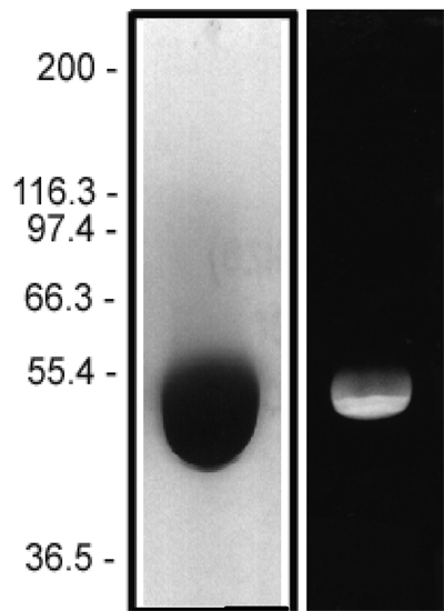
Figure 1: HiLyte Fluor™ 647 tubulin protein purity determination. A 50 µg sample of unlabeled tubulin protein was separated by electrophoresis in a 4-20% SDS-PAGE system and stained with Coomassie Blue (A). Protein quantitation was performed using the Precision Red Protein Assay Reagent (Cat. # ADV02). 20 µg of the same protein sample was run in a 4-20% SDS-PAGE system and photographed directly under 525-625nm illumination (B).
Biological Activity
The biological activity of HiLyte Fluor™ 647 tubulin is assessed by a tubulin polymerization assay. To pass quality control, a 5 mg/ml solution of HiLyte Fluor™ 647 labeled tubulin in G-PEM plus 5% glycerol must polymerize to >85%. This is comparable to unlabeled tubulin under identical conditions.
Related Products
Related Products
Tubulin Protein (>99% Pure): Porcine Brain | T240
 | LMN02")
 | LMN02")
Laminin (Green Fluorescent, HiLyte 488) | LMN02

Tubulin Polymerization Assay Using >99% Pure Tubulin, Fluorescence Based | BK011P
, His-Tag")
Porcine Circovirus 2 Capsid Protein (E. coli), His-Tag
")