
Laminin (Green Fluorescent, HiLyte 488) | LMN02
 | LMN02")
- SKU:
- LMN02-A | LMN02-B
- Availability:
- In Stock
 | LMN02")
 | LMN02")
 | LMN02")
 | LMN02")
Description
Laminin (Green Fluorescent, HiLyte 488) | LMN02
Product Uses Include
- Cell invasion assays (1)
- FACS analysis of laminin binding cells (2)
Material
Laminin-1 is purified from EHS tumor tissue and is free of the laminin binding protein entactin which is a common contaminant in some laminin preparations (150 kDa). Protein purity is determined by scanning densitometry of Coomassie Blue stained protein on a 4-20% polyacrylamide gel. The laminin is >90% pure (Figure 1).
The protein is modified to contain covalently linked HiLyte 488TM dyes (3) at random surface lysines. An activated ester of HiLyte 488TM is used to label the protein. Labeling stoichiometry is determined by spectroscopic measurement of protein and dye concentrations. Final labeling stoichiometry is 2-5 dyes per protein molecule (Figure 2). The material is guaranteed to contain <15% of free dye and >85% of dye conjugated to laminin. HiLyte 488TM laminin can be detected using a filter set of 502nm excitation and 527nm emission.
Laminin runs as individual subunits on SDS-PAGE with an apparent molecular weight of 400 and 225 kDa (Figure 1). LMN02 is supplied as an off white lyophilized powder. Each vial of LMN02 contains 20 µg protein.
Purity
Purity is determined by scanning densitometry of proteins on SDS-PAGE gels. Samples are >90% pure.
Figure 1: HiLyte 488TM Laminin Purity Determination
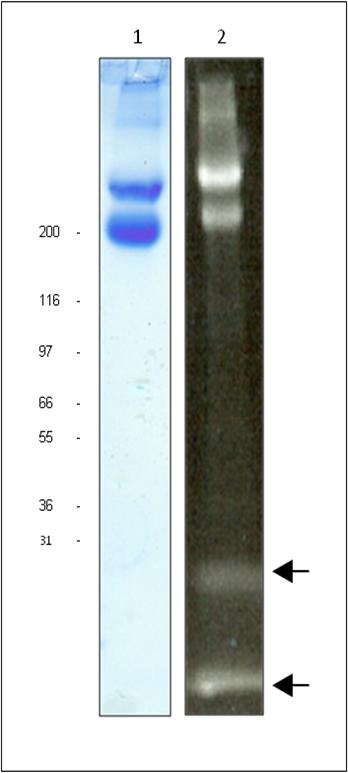
Legend: 20 µg of unlabeled laminin (Lane 1) and 20 µg of HiLyte 488TM laminin (Lane 2) was separated by electrophoresis in a 4-20% SDS-PAGE system. The unlabeled protein was stained with Coomassie Blue and visualized in white light. The labeled protein was visualized under UV light. The alpha subunit runs at 400 kDa (top band) while the beta and gamma subunits run as a 225 kDa doublet (lower band). Arrows indicate unincorporated dye. In this example unincorporated dye = 13%. Protein quantitation was determined with the Precision Red™ Protein Assay Reagent (Cat. # ADV02). Mark12 molecular weight markers are from Invitrogen.
Figure 2: Absorption scan of HiLyte 488TM laminin in solution
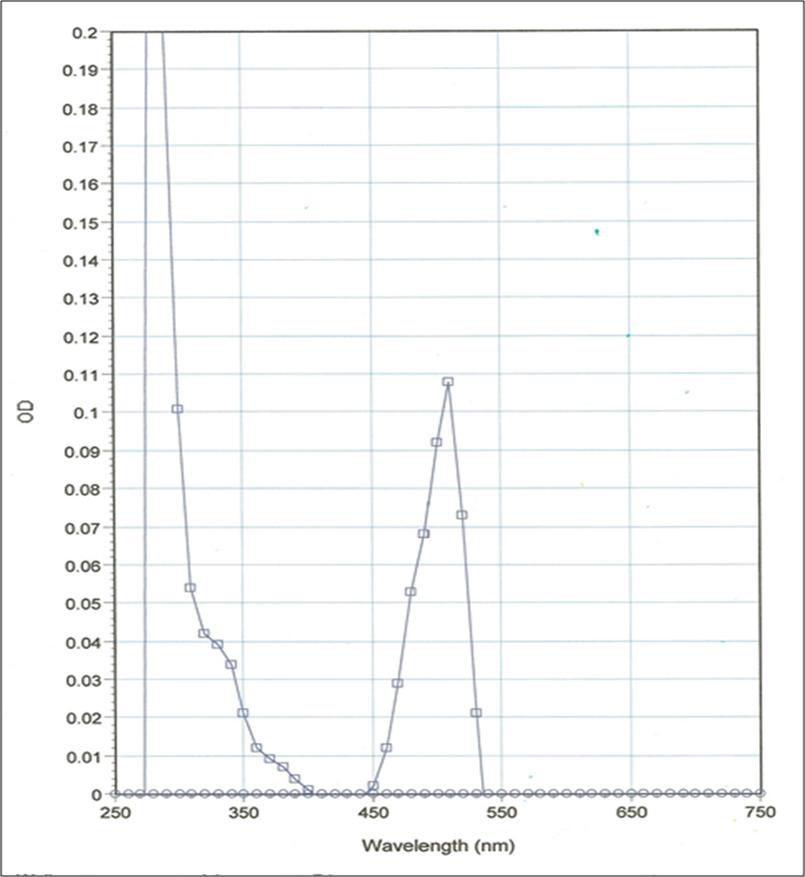
Legend: LMN02 was diluted with Milli-Q water and its absorbance spectrum was scanned between 250 and 750 nm. In this example, HiLyte 488TM labeling stoichiometry was calculated to be 3.5 dyes per laminin protein using the absorbancy maximum for HiLyte 488TM at 527 nm and the Beer-Lambert law. Dye extinction coefficient when protein bound is 70,000M-1cm-1 .
Storage and Reconstitution
Shipped at ambient temperature. The lyophilized protein can be stored desiccated to <10% humidity at 4°C for 6 months in the dark. For reconstitution, briefly centrifuge to collect the product at the bottom of the tube and resuspend to 1 mg/ml with 20 µl cold distilled water. The protein will then be in the following buffer: 100 mM PIPES pH 7.2, 1% dextran and 5% (w/v) sucrose. Avoid excessive mixing as this can cause protein aggregation. The concentrated protein should be aliquoted into experiment sized amounts, snap frozen in liquid nitrogen and stored at –70°C where it is stable for 6 months. For working concentrations, further dilution of the fluorescent laminin should be made in a suitable buffer or tissue culture media. HiLyte 488TM laminin is a labile protein and should be handled with care. Avoid repeated freezethaw cycles.
Related Products
Related Products
: Porcine Brain | TL670M")
: Porcine Brain | TL670M")
Tubulin Protein (Fluorescent HiLyte 647): Porcine Brain | TL670M
")
iMatrix-511(recombinant laminin-511)
")
Live Cell Tracking Kit (Green Fluorescence) | KTA1002
")
TUNEL Apoptosis Detection Kit (Green Fluorescence) | KTA2010
")