Description
C-FOS Antibody | 26209 | ImmunoStar
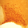
For induction of c-fos protein activity rats were injected with 1.0 ml of 1.5 M NaCl per 100 grams of body weight. Negative control rats were injected with the same volume of normal saline. The ImmunoStar c-fos antiserum was quality control tested using standard immunohistochemical methods. The antiserum demonstrates strongly positive labeling of rat paraventricular nucleus and supraoptic nucleus using indirect immunofluorescent and biotin/avidin-HRP techniques. No labeling was seen in negative control rats. Recommended primary dilutions for these methods are 1/4000-1/6000 in PBS/0.3% Triton X-100 – FITC Technique and 1/4000-1/6000 in PBS/0.3% Triton X-100 – biotin/avidin-HRP Technique. Injection of intraperitoneal hypertonic saline to provoke fos expression is most usually used to set ICC staining parameters. It is critical that tissue is harvested 60-90 minutes post injection for this and any other stress that is used. Inject 1 ml/100 g body weight 1.5 M NaCl intraperitoneally. Harvest brains 90 minutes post injection. Time is critical. Specificity of the antiserum was demonstrated by blockage of staining in experimental rats by omission of c-fos antibody or by substitution of antibody pre-incubated with synthetic peptide or the conjugate. Immunoblot analysis of mediobasal hypothalamus showed a single band of approximately 55-60 kD.Host: Rabbit
State: Lyophilized Whole Serum
Reacts With: Mouse, Rat
Availability: In Stock
Alternate Names: Proto-oncogene c-Fos; Cellular oncogene; G0/G1 switch regulatory protein 7; Proto-oncogene protein; G0S7; p55; AP-1; FBJ murine osteosarcoma viral oncogene homolog, anti-CFOS
Gene Symbol: FOS
RRID: AB_572267


