
The Guide to Flow Cytometry Calibration Beads
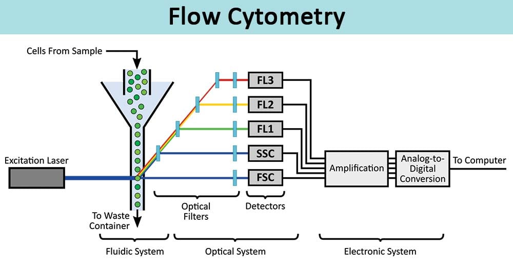
Flow cytometry is one of the most powerful techniques in modern life sciences, enabling precise cell analysis at high speed. But even the most advanced cytometer can produce unreliable data without proper calibration.
This is where calibration beads (fluorescent particles) become essential.
In this guide, you’ll discover:
- What calibration beads are?
- How they work in flow cytometry?
- Why they are critical for quality control (QC)?
- How to use them effectively?
- Best practices to ensure reproducible results.
What Are Flow Cytometry Calibration Beads?
Calibration beads are microspheres embedded with fluorescent dyes used to standardize and validate the performance of flow cytometers.
They are designed to:
- Emit fluorescence across multiple wavelengths
- Provide consistent signal intensity
- Mimic biological particles in size and optical behavior
These particles allow researchers to monitor instrument performance over time and ensure data consistency across experiments.
=> According to the document, these particles are widely used for :
- Routine calibration.
- Long-term performance tracking.
- Instrument alignment.
- Fluorescence microscopy validation.
How Do Multi-Fluorescent Calibration Beads Work?
Modern calibration beads contain multiple fluorophores within a single particle, enabling detection across different channels.
Key mechanism :
- Each bead population emits different fluorescence intensities.
- These intensities appear as distinct peaks in histograms.
- Each peak corresponds to a specific fluorescence level.
Why Calibration Beads Are Critical in Flow Cytometry?
1. Ensure Data Accuracy
Without calibration, fluorescence signals can drift, leading to incorrect biological conclusions.
2. Enable Reproducibility
Calibration beads provide standardized reference points, making results comparable across :
- Different days.
- Different instruments.
- Different laboratories.
3. Monitor Instrument Performance
They help detect:
- Laser instability.
- Detector sensitivity loss.
- Alignment issues.
4. Define Dynamic Range
Calibration beads allow users to determine the operational range of detectors, especially photomultiplier tubes (PMTs).
Key Features of High-Quality Calibration Beads
The most effective calibration particles share these characteristics:
✔ Multi-channel compatibility
- Excitable from 365 to 650 nm
- Suitable for most cytometer configurations
✔ Multiple intensity levels
- Different fluorescence peaks on same-size beads
- Enables full detector range calibration
✔ High stability
- Fluorophores are embedded inside particles
- Resistant to photobleaching
✔ Freeze-thaw resistance
- Can be stored and reused without signal degradation
✔ Chemical resilience
- Can be sanitized (e.g., ethanol treatment)
=> These features ensure long-term reliability and cost efficiency.
Understanding Calibration Curves & Linearity
One of the most important applications is generating calibration curves.
These curves :
- Plot fluorescence intensity vs detector response.
- Validate linearity of the cytometer.
- Ensure accurate quantification.
=> The calibration graphs demonstrate how fluorescence intensity correlates with detector channels, confirming instrument linearity.
How to Use Calibration Beads (Step-by-Step)?
Step 1: Prepare Dilution
- Add a few drops of bead suspension to ~1 mL buffer.
Step 2: Run on Cytometer
- Acquire data using standard settings.
Step 3: Identify Peaks
- Detect distinct fluorescence populations.
Step 4: Adjust Settings
- Optimize voltage and gain.
- Ensure peaks are correctly spaced.
Step 5: Record & Track
- Store results for QC monitoring.