Description
Glycosaminoglycans Assay Kit - Cat Number: 6022 From Chondrex.
Research Field: ECM
Clonality: N/A
Cross-Reactivity:
Host Origin: N/A
Applications: N/A
Isotype: N/A
Detection Range: 50 µg/ml-3.1µg/ml
Sample Type: Tissue Extract, Cultured Cells
Concentration: N/A
Immunogen:
PRODUCT SPECIFICATIONS
DESCRIPTION: Assay kit to quantify GAGs
FORMAT: 96-well ELISA plate with removeable strips
ASSAY TYPE: Colorimetric assay
ASSAY TIME: 5 minutes
STANDARD RANGE: 50 µg/ml to 3.1 µg/ml
NUMBER OF SAMPLES: Samples NOT containing extra proteins: up to 42 (duplicate) samples/plate
Samples containing extra proteins: up to 21 (duplicate) samples/plate
SAMPLE TYPES: Tissue homogenate
RECOMMENDED SAMPLE DILUTIONS: Varies
CHROMOGEN: N/A (read at 525 nm)
STORAGE: -20°C for 12 months
VALIDATION DATA: Intra-Assay (1.7-7.6%)/Inter-Assay (5.2-8%)/Spiking Test (106-117%)
NOTES: This assay uses a 5-point standard curve
Samples need to be solubilized before running assay
INTRODUCTION
Glycosaminoglycans (GAGs) are negatively charged polysaccharides located in most connective tissues and extracellular matrices (ECM),
as well as on the surfaces of many cell types. Consisting of repeating core disaccharide units, GAGs are categorized into four types:
heparan/heparan sulfate, chondroitin/dermatan sulfate, keratin sulfate, and non-sulfated hyaluronan. Sulfated GAGs in the ECM exist as
proteoglycans which typically consist of multiple glycosaminoglycan chains attached to a core protein (1). In a highly organized ECM, articular
cartilage is composed of type II collagen, hyaluronan, link protein, and chondroitin sulfate-rich proteoglycans, which provide the osmotic
resistance necessary for cartilage to resist compressive loads (2).
In addition to type II collagen, GAGs are considered an autoantigen of rheumatoid arthritis (RA), as anti-GAGs antibodies associated with
ECM degradation exist in the serum of RA patients (3). Although the role of proteoglycans in RA is still unknown, research shows that
immunizing mice with proteoglycans consisting of GAGs can induce arthritis (4). Similarly, the loss of ECM is the first observable change in
osteoarthritis (OA), therefore, determining GAGs levels may be a useful marker of disease progression. Furthermore, the successful use of
artificial cartilage for the treatment of OA necessitates the analysis of type II collagen and GAGs content to ensure the cartilage quality (5).
Chondrex, Inc. provides a sulfated GAGs Assay Kit (Cat # 6022) using cationic dye 1,9 dimethylmethylene blue (DMB) which binds to highly
charged sulfated GAGs, not including hyaluronan (6). This kit utilizes an improved DMB solution, minimizing interference with negatively
charged contaminants such as DNA and RNA and uses chondroitin sulfate as a standard for the analysis of ECM in cartilage. Moreover, to
analyze collagen coexisting with GAGs in cartilage, Chondrex, Inc. provides a variety of type and species-specific collagen detection kits
and total collagen detection kits. For more information, please visit www.chondrex.com or contact us at support@chondrex.com.
KIT COMPONENTS

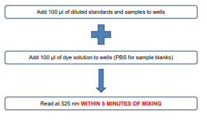
ASSAY OUTLINE

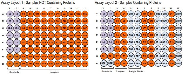
PLATE MAPPING

NOTES BEFORE USING ASSAY
NOTE 1: It is recommended that the standard and samples be run in duplicate.
NOTE 2: Warm up all buffers to room temperature before use.
NOTE 3: Crystals may form in Wash Buffer, 20X when stored at cold temperatures. If crystals have formed, warm the wash buffer by placing
the bottle in warm water until crystals are completely dissolved.
NOTE 4: Measure exact volume of buffers using a serological pipet, as extra buffer is provided.
NOTE 5: Cover the plate with plastic wrap or a plate sealer after each step to prevent evaporation from the outside wells of the plate.
NOTE 6: Samples need to be solubilized. Please inquire at support@chondrex.com for Chondrex, Inc.’s “Tips on Glycosaminoglycan
Solubilization”
NOTE 7: Guanidine used for extracting GAGs from samples will interfere with this assay. Guanidine extracted samples should be diluted or
dialyzed against PBS to reduce the guanidine concentration to less than 0.25 M. In addition, some proteins have an absorbance at 525
nm; therefore, samples contaminated with unnecessary proteins must be diluted to a 5% or less protein solution and require sample blanks
to ensure accurate results. Please refer to assay layout 2 for samples containing proteins.


