Description
Bacterial Collagenase Assay Kit - Cat Number: 3014 From Chondrex.
Research Field: ECM
Clonality: N/A
Cross-Reactivity:
Host Origin: N/A
Applications: N/A
Isotype: N/A
Detection Range: Detection Limit of 0.5 units
Sample Type: Culture Media, Tissue Homogenate
Concentration: N/A
Immunogen:
PRODUCT SPECIFICATIONS
DESCRIPTION: Assay kit to assess collagenase activity and inhibitory activity to collagenase
FORMAT: 96-well ELISA plate with non-removeable strips
ASSAY TYPE: Enzyme Assay/Fluorescence-based Assay
ASSAY TIME: Approximately 2 hours
STANDARD RANGE: Depends on incubation time
NUMBER OF SAMPLES: Activity Assay: Up to 41 (duplicate) samples/plate
Inhibitor Assay: Up to 45 (duplicate) samples/plate
SAMPLE TYPES: Culture Media and Tissue Homogenate
RECOMMENDED SAMPLE DILUTIONS: Depends on enzyme activity in samples
CHROMOGEN: N/A (Read Fluorescence Intensity at Emission 520 nm/Excitation 490 nm)
STORAGE: -20°C for 12 months
VALIDATION DATA: N/A
INTRODUCTION
Collagenase produced by Clostridium histolyticum, isolated by Mandl et al. in 1953 (1), has been widely used in research and clinical fields
for isolating cells and digesting connective tissues (2). Importantly, bacterial collagenases differ from mammalian collagenases in their
substrate specificity, as bacterial collagenases cleave the native collagen molecule into multiple fragments, whereas mammalian
collagenases cleave the collagen molecule at a single site into two fragments. Clostridium species infection causes severe tissue necrosis
leading to gas gangrene (3). At the infection site, collagenases from Clostridium species facilitate extensive unregulated destruction of the
extracellular matrix because of the absence of bacterial collagenase inhibitors in animal and human sera, whereas mammalian collagenase
activity is strictly regulated by mammalian serum inhibitors (4-6).
Chondrex, Inc. provides a rapid bacterial collagenase assay kit using soluble FITC-labeled type I collagen as a substrate instead of radiolabeled collagen (7). This kit can be used not only for assaying collagenase activity, but also for inhibitor assay and includes protocols for
both.
Note: The reference collagenase provided in this kit is a positive control, not a standard. Collagenase activity in samples should be
determined based on the amounts of collagen substrate digested.
KIT COMPONENTS

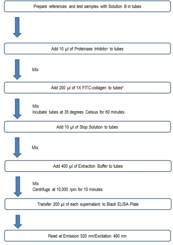
ASSAY OUTLINE (FOR BOTH COLLAGENASE ACTIVITY AND INHIBITOR ASSAY)

ASSAY PROCEDURE: COLLAGENASE ACTIVITY ASSAY
1. Prepare Microcentrifuge Tubes and Reference: Prepare amber 1.5 ml microcentrifuge tubes for Buffer, 100% Control, Blank,
Reference Collagenases, and Test Samples as shown on the Collagenase Activity assay sheet. Dissolve one vial of Reference
collagenase in 1 ml of Solution B. Partially used Reference collagenase stock solution can be kept at - 20°C for future assays.
NOTE: Proteins in sample specimens may cause quenching, and consequently, fluorescent intensity (FI) determined in sample tubes
might be underestimated. For example, if the collagenase activity is very low in a sample solution which contains a certain amount of
contaminant proteins, the FI in the samples will be lower than the Blank value. In order to correct these under-estimated results, the
identical sample mixed with Stop Solution should be added to the Blank tubes and 100% Control tubes. This quenching is mainly caused
by turbidity formed by the proteins in the Extraction Buffer. Similarly, colors or dyes in bacteria culture media also causes quenching. In
this case, add the same culture media to Blank and 100% Control tubes.
2. Add References and Samples: Add the proper amounts of Solution B, Reference Collagenase, and test samples to tubes and adjust
the final volume to 190 µl as shown on the assay sheet. The buffer tube should have 390 µl of Solution B.
NOTE 1: Because bacterial collagenase exists as an active form, collagenase activation is not necessary.
NOTE 2: The test sample volumes may vary from 1 -100 µl. However, the final assay sample volume should be adjusted to 190 µl with
Solution B.
3. Add Proteinase Inhibitor: Dissolve one vial of proteinase inhibitor in 1 ml of Solution B. Add 10 µl of proteinase inhibitor into all tubes
to neutralize non-collagenolytic proteinases in solution.
4. Prepare 1X FITC: Prepare a 1X FITC-collagen solution by mixing an equal volume of the 2X FITC-collagen and cold Solution A (200
µl of the mixture is required for each sample to be tested) in a container protected from light, such as an amber colored tube or bottle
(FITC is light sensitive).
5. Add 1X FITC: Add 200 µl of the 1X FITC-collagen solution into all tubes (200 µl) except for the Buffer tube. Mix well and incubate at
35°C for 10-120 minutes.
NOTE: Incubate the reference and 100% control tubes for 60 minutes at 35°C. The incubation time for test samples will vary depending
on the collagenase activity in sample specimens. Do not incubate test samples longer than 120 minutes otherwise they may yield high
background levels. Background refers to the degradation of collagen due to extended exposure to high temperatures.
6. Add Stop Solution: Stop the collagenase reaction by adding 10 µl of Stop Solution to each tube and mixing well.
7. Add Extraction Buffer: Cool samples to room temperature. Add 400 µl of Extraction Buffer to each tube. Do not use cold buffer. Mix
vigorously and centrifuge at 10,000 rpm for 10 minutes.
8. Transfer: Carefully transfer 200 µl of each supernatant (in duplicate) into the black 96-well plates provided in the kit and determine the
fluorescence intensity (FI) at λEm = 520 nm and λEx = 490 nm.
NOTE: Supernatants contaminated with pellets in the 96-well plate will lead to high FI values, resulting in overestimated assay results
NOTES: Uses FITC
1 Review
-
Original
Gentaur provides original things to their customers and never found any complaints about their products


