
Rapid Collagenase Assay Kit with Type I Collagen Substrate

- SKU:
- 445-3001
- Size:
- 1 kit x2 plates
- Shipping:
- Dry Ice
- Storage:
- -80 C

Description
Rapid Collagenase Assay Kit with Type I Collagen Substrate - Cat Number: 3001 From Chondrex.
Research Field: ECM
Clonality: N/A
Cross-Reactivity:
Host Origin: N/A
Applications: N/A
Isotype: N/A
Detection Range: Detection Limit of 0.5 units
Sample Type: Culture Media, Tissue Homogenate
Concentration: N/A
Immunogen:
PRODUCT SPECIFICATIONS
DESCRIPTION: Assay kit to assess collagenase activity
3001: Rapid Collagenase Assay Kit with Type I Collagen Substrate
3002: Rapid Collagenase Assay Kit with Type II Collagen Substrate
FORMAT: 96-well ELISA Plate with non-removeable strips
ASSAY TYPE: Enzyme Assay/Fluorescence-based Assay
ASSAY TIME: 2.5 hours
STANDARD RANGE: Depends on incubation time
NUMBER OF SAMPLES: Up to 44 (duplicate) samples/plate
SAMPLE TYPES: Culture Media and Tissue Homogenate
RECOMMENDED SAMPLE DILUTIONS: Depends on enzyme activity in samples
CHROMOGEN: N/A (Read at Emission 520 nm/Excitation 490 nm)
STORAGE: -20°C for 12 months (Reference Standard is stored at -80°C)
VALIDATION DATA: N/A
NOTES: Uses FITC
INTRODUCTION
Collagenases are members of the matrix metalloproteinase (MMP) family and degrade collagen types I, II, and III. At least three distinct forms of collagenase (MMP-1, MMP-8, and MMP-13) have been identified. Collagenases are produced by many types of cells such as myeloid and fibrosarcoma cells. Increased collagenase levels have been found in physiological conditions, such as post-partum uterine tissue or tadpole metamorphosis, and pathological conditions, such as inflammation and tumor metastasis. These collagenases have almost identical substrate specificities. However, individual collagenases may have unique enzyme-substrate affinities, resulting in different physiological and pathological roles in the turnover of collagen depending on the tissues and cell types. For example, MMP-13 digests type II collagen ten times faster than type I collagen.
This kit is designed to assay mammalian collagenase activities in approximately two hours using FITC-labeled telopeptide-free soluble bovine type I collagen (Catalog # 3001) or bovine type II collagen (Catalog # 3002) as a substrate (1). This kit only requires 1/10 of the assay time of conventional assay methods that use a collagen gel substrate and it also has higher assay sensitivity than assays using radioisotopelabeled collagen (2).
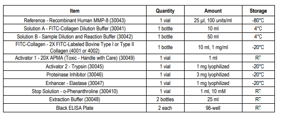
KIT COMPONENTS
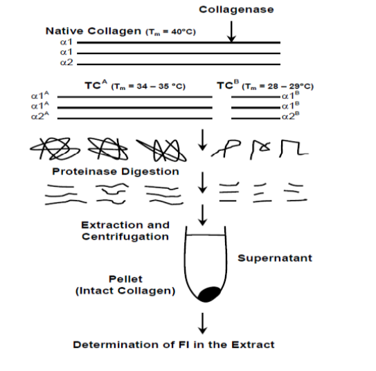
PRINCIPLE OF THE RAPID COLLAGENASE ASSAY KIT (TYPE I COLLAGEN)
Mammalian collagenases cleave the alpha chain triple helixes of collagen, yielding 3/4 and 1/4 collagen fragments, TCA and TCB fragments.
The denaturation temperature of these fragments is 34-35°C and 28-29°C respectively, whereas the denaturation temperature of intact collagen is 40°C. Therefore, these cleaved fragments selectively denature into single random coils at 35°C which can be extracted with Extraction Buffer. To shorten the denaturation process, which normally takes 60 minutes, an enhancer is used to further digest collagenasedegraded products into small peptides (2).
NOTES BEFORE USING ASSAY
NOTE 1: In general, collagenases are secreted as latent proenzymes and require proteolytic conversion for activation. However, collagenase activity is strictly regulated by tissue inhibitors of metalloproteinase (TIMP) and by 2 macroglobulins (2M) in serum. Thus, different conditions are required to activate collagenase depending on the experimental purpose, source of enzyme, and levels of proteinase inhibitors in individual specimens.
NOTE 2: APMA (4-aminophenylmercuric acetate) is widely used to activate latent pro-collagenase (3). Activator 1 (APMA) contains mercury which is very toxic if inhaled, ingested, or makes contact with skin. Neurological hazard target organs include the kidneys and nerves. Wear suitable protective gloves, clothing, and eyewear.
NOTE 3: Trypsin activates both latent pro-collagenases and collagenases bound by inhibitors such as 2M and low molecular weight collagenase inhibitors (4, 5). However, soybean trypsin inhibitor (SBTI) must be added to neutralize the added trypsin before assaying
collagenase activity.
NOTE 4: Potassium thiocyanate (KSCN) or potassium iodide (KI) reactivates collagenases bound by inhibitors such as 2M (6). Another activation method to consider is to dialyze samples against 3M KSCN dissolved in 0.05M Tris-HCl buffer, pH 7.5, at 4°C overnight. Then, remove the KSCN by dialyzing against 0.05M Tris-HCl buffer, pH 7.8, containing 0.2M NaCl and 5mM CaCl2. These reagents may be useful for denaturing collagenase inhibitors in sample specimens prior to activating pro-collagenase by APMA (7).
NOTE 5: Both dithiothreitol (DTT) and iodoacetamide have been reported to reactivate collagenases bound by TIMPs (8). However, these reagents also may inactivate collagenase by reducing the disulfide bonds and alkylating glutamic acid at the active site. Therefore, the limitations of this method must be taken into consideration.
Related Products
Related Products

Rapid Collagenase Assay Kit with Type II Collagen Substrate
 Assay Kit")
Type I Collagen C-Telopeptide (CTX-I) Assay Kit

FITC-Labeled Bovine Type I Collagen Substrate

Bacterial Collagenase Assay Kit
