
Mouse Anti-OVA IgG1 Antibody Assay Kit

- SKU:
- 445-3013
- Size:
- 1 kit
- Shipping:
- Gel Packs
- Storage:
- -20 C

Description
Mouse Anti-OVA IgG1 Antibody Assay Kit - Cat Number: 3013 From Chondrex.
Research Field: Allergy, Immunology
Clonality: N/A
Cross-Reactivity:
Host Origin: N/A
Applications: N/A
Isotype: N/A
Detection Range: 12.5 ng/ml-0.2 ng/ml
Sample Type: Serum, Plasma
Concentration: N/A
Immunogen:
DESCRIPTION: ELISA kit to quantify mouse anti-ovalbumin antibodies
FORMAT: 96-well ELISA Plate with removeable strips
ASSAY TYPE: Indirect ELISA
ASSAY TIME: 4.5 hours
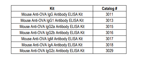
STANDARD RANGE: 3011 (IgG) : 12.5 - 0.2 ng/ml
3013 (IgG1) : 12.5 - 0.2 ng/ml
3015 (IgG2a) : 50 - 0.8 ng/ml
3016 (IgG2b) : 50 - 0.8 ng/ml
3017 (IgM) : 100 - 1.6 ng/ml
3018 (IgA) : 12.5 - 0.2 ng/ml
3029 (IgG2c) : 50 - 0.8 ng/ml
NUMBER OF SAMPLES: Up to 40 (duplicate) samples/plate
SAMPLE TYPES: Serum & Plasma
RECOMMENDED SAMPLE DILUTIONS: 1:100 (at least)
CHROMOGEN: TMB (read at 450 nm)
STORAGE: -20°C for 12 months
VALIDATION DATA: 3011: Intra-Assay (2.6-9.5%)/Inter-Assay (0.4-3.7%)/Spiking Test (93-107%)
3013: Intra-Assay (4.7-9.1%)/Inter-Assay (3.6-9.5 %)/Spiking Test (96-114%)
3015: Intra-Assay (5.9-8.9%)/Inter-Assay (6.3-9.4%)/Spiking Test (98-104%)
3016: Intra-Assay (3.9-9.9%)/Inter-Assay (6.3-9.4%)/Spiking Test (94-106%)
3017: Intra-Assay (2.9-4.6%)/Inter-Assay (6.6-9.1%)/Spiking Test (93-116%)
3018: Intra-Assay (0.2-7.8%)/Inter-Assay (1.2-7.9%)/Spiking Test (93-98%)
3029: Intra-Assay (5.5-9.6%)/Inter-Assay (2.9-7%)/Spiking Test (100-109%)
INTRODUCTION
Ovalbumin (OVA) is a widely used antigen for inducing allergic reactions in experimental animals (1-4). To study the contribution of antibodies to allergic reactions, Chondrex, Inc. provides a variety of subtype and subclass anti-OVA mouse antibody ELISA kits. Investigators studying the pathogenesis of allergic diseases in mice should consider several issues such as mucosal immunity, antibody responses to different subtypes ( IgA, IgE, IgG, and IgM) and subclasses (IgG1, IgG2a, IgG2b, and IgG2c), and even the types of adjuvants used for immunization. The following may be informative for studies on allergic reactions in experimental animals. 1) Mucosal immunity: The mucosal immune system is the first line of defense against potential pathogenic and non-pathogenic environmental factors such as bacteria, viruses, and dietary proteins. Importantly, poor mucosal immune function may lead to abnormal absorption of mimic antigens such as food components and bacterial cell walls, which elicit antibodies that may cross-react with autologous components, also known as “autoantibodies”. IgG, the dominant immunoglobulin subtype in serum, protects the host from pathogens and unwanted antigens that have penetrated the body. On the other hand, IgA, a known mucosal immunoglobulin, preventsthe penetration of pathogens and other unwanted antigenic substances through mucosal membranes. Therefore, to study mucosal and systemic immune responses to allergens in mouse models, OVA is a valuable and convenient antigen (5). 2) Allergy: In general, an allergic reaction is mediated by IgE-antigen complexes. More specifically, IgE molecules cross-linked by a polyvalent antigen on the surfaces of mast cells trigger their degranulation which initiates the ensuing allergic cascade. Although the role
of IgG antibodies in allergic reactions is not yet clear, two opposing roles are postulated: 1) IgG antibodies which share epitopes with IgE antibodies may competitively bind the epitopes on the allergen and modulate the allergic reaction, or 2) IgG antibodies may enhance the allergic reaction by providing aggregated allergens to IgE on mast cells. In addition, the roles of antibody subtypes and subclasses may differ depending on the allergic reaction, as IgG1 and IgE are regulated by Th2 cells, whereas IgG2a, IgG2b and IgG2c are dependent on Th1 cells. Thus, to investigate the immune responses involved in allergic reactions in OVA-induced allergic mouse models (1-4), antiOVA IgE, IgG, and IgA antibody ELISA kits are valuable tools.
3) Adjuvants: The type of adjuvant used can elicit specific antibody subtypes. For example, aluminum adjuvant is widely used to elicit IgE antibodies (7), whereas Cholera toxins are effective at eliciting IgA antibodies (6). Moreover, Complete Freund’s Adjuvant (CFA) is widely used for stimulating IgG and IgM antibody production (8).
LIST OF MOUSE ANTI-OVA ANTIBODY SUBTYPE/SUBCLASS ELISA KITS
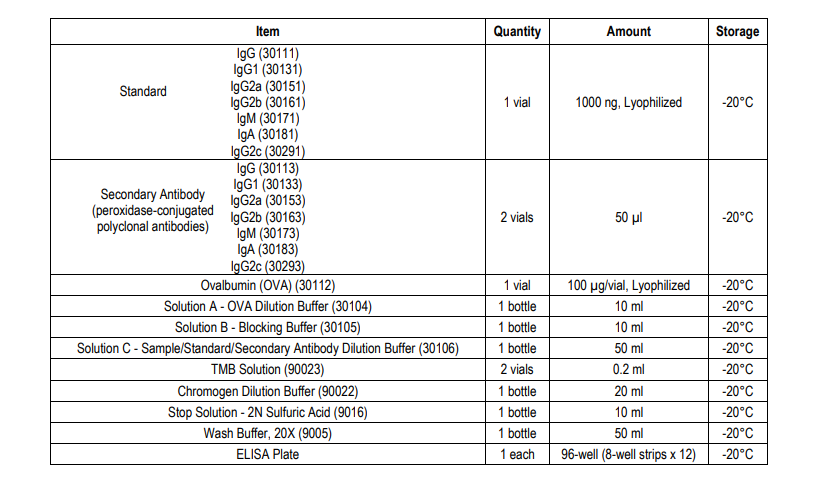
KIT COMPONENTS
NOTES BEFORE USING ASSAY
NOTE 1: It is recommended that the standard and samples be run in duplicate.
NOTE 2: Warm up all buffers to room temperature before use.
NOTE 3: Crystals may form in Wash Buffer, 20X when stored at cold temperatures. If crystals have formed, warm the wash buffer by placing the bottle in warm water until crystals are completely dissolved.
NOTE 4: Measure exact volume of buffers using a serological pipet, as extra buffer is provided.
NOTE 5: Cover the plate with plastic wrap or a plate sealer after each step to prevent evaporation from the outside wells of the plate.
NOTE 6: For partial reagent use, please see the assay protocol’s corresponding step for the appropriate dilution ratio. For example, if the protocol dilutes 50 µl of a stock solution in 10 ml of buffer for 12 strips, then for 6 strips, dilute 25 µl of the stock solution in 5 ml of buffer. Partially used stock reagents may be kept in their original vials and stored at -20⁰C for use in a future assay.
NOTE 7: This kit contains animal components from non-infectious animals and should be treated as potential biohazards in use and for disposal.
NOTE 8: Depending on the isotypes, subtypes, and targeting epitopes of antibodies, the binding affinity of individual antibodies varies significantly. Therefore, the total IgG antibody concentration calculated as the sum of individual IgG subtypes might not perfectly match the total IgG concentration as determined by the Mouse Anti-OVA IgG Antibody ELISA Kit (Cat # 3011)
ASSAY OUTLINE
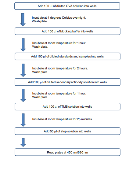
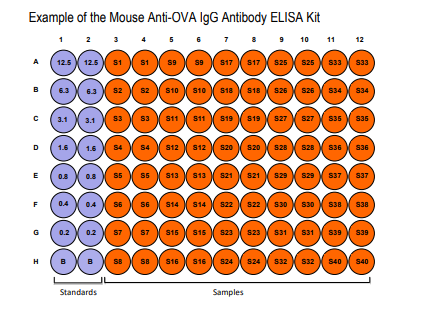
PLATE MAPPING
ASSAY PROCEDURE
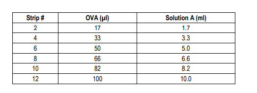
Add OVA Solution: Dissolve one vial of Ovalbumin (OVA) with 10 ml of OVA Dilution Buffer (Solution A). Alternatively, dissolve one vial of OVA with 100 µl of Solution A and dilute according to the table below. Add 100 µl of OVA solution to each well and incubate at 4°C overnight.
2. Wash: Dilute 50 ml of 20X wash buffer in 950 ml of distilled water (1X wash buffer). Wash the plate with 1X wash buffer at least 3 times using a wash bottle with manifold or an automated plate washer. Empty the plate by inverting it and blotting on a paper towel to remove excess liquid. Do not allow the plate to dry out. 3. Add Blocking Buffer: Add 100 µl of the Blocking Buffer (Solution B) to each well and incubate at room temperature for 1 hour.
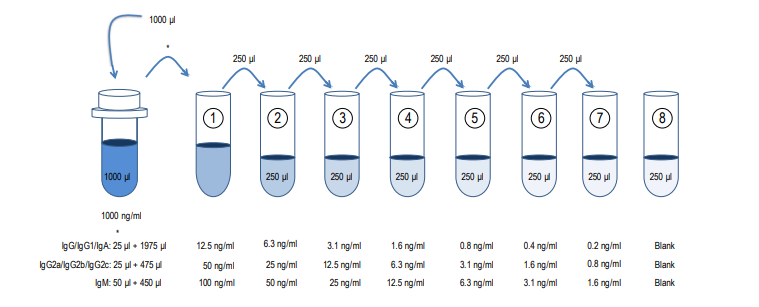
4. Prepare Standard Dilutions: Please see the figure below for each assay’s recommended standard range. Dissolve one vial of Standard in 1 ml of Sample/Standard/Secondary Antibody Dilution Buffer (Solution C) and keep it as a standard stock. Dilute the standard stock accordingly to get the first stock solution. Then serially dilute it with Solution C. For example, mix 250 µl of the first stock solution with an equal volume of Solution C to make the second stock solution, and then repeat it five more times. The original standard stock solution may be stored at -20⁰C for use in a future assay. Chondrex, Inc. recommends making fresh serial dilutions for each assay.
Prepare Sample Dilutions: The dilution of serum from mouse immunized with OVA varies (1:100 or more) depending on the
immunization schedule and timing of serum collection. In general, no antibodies against OVA are observed in normal serum at a 1:100 dilution. 6. Wash: Wash the plate with 1X wash buffer at least 3 times using a wash bottle with manifold or an automated plate washer. Empty the plate by inverting it and blotting on a paper towel to rem
Related Products
Related Products

Mouse Anti-OVA IgM Antibody Assay Kit

Mouse Anti-OVA IgG2b Antibody Assay Kit

Mouse Anti-OVA IgG2c Antibody Assay Kit

Mouse Anti-OVA IgG Antibody Assay Kit
