
FITC-Dextran, 40 kDa, 25 mg/ml x 5 ml

- SKU:
- 445-4009
- Size:
- 25 mg/ml x 5 ml
- Shipping:
- Gel Packs
- Storage:
- 4 C

Description
FITC-Dextran, 40 kDa, 25 mg/ml x 5 ml - Cat Number: 4009 From Chondrex.
Research Field: Colitis, Membrane Permeability
Clonality: N/A
Cross-Reactivity:
Host Origin: N/A
Applications: N/A
Isotype: N/A
Detection Range: N/A
Sample Type: N/A
Concentration: 25 mg/ml
Immunogen:
PRODUCT SPECIFICATIONS
DESCRIPTION: Fluorescein isothiocyanate (FITC) labeled dextran
APPLICATION: Use to assess the permeability of semi-permeable membranes either in vivo or in vitro (1-4).
NOTE: FITC-dextran can be used simultaneously with TRITC-dextran (Cat # 4014) as
fluorescence occurs at different wavelengths.
QUANTITY: 5 ml
FORM: 25 mg/ml solution in 0.05M phosphate buffered saline
MOLECULAR WEIGHT: 40 kDa
FLUORESCENCE: Excitation: 490 nm, Emission: 520 nm
STORAGE: 4⁰C in the dark
STABILITY: 1 year
IN VIVO PROTOCOL:
1. Fast mice 4 hours before oral feeding and for the duration of the experiment.
2. Feed 20 ml/kg by oral gavage.
3. Maintain fasting conditions and wait 3 hours (may vary depending on individual animals).
4. Collect blood by retro-orbital bleeding, then spin and collect plasma. Dilute plasma 1:2 (or
more) with PBS.
NOTE 1: Protein in the samples may interfere with and reduce the fluorescence intensity
(FI). Therefore, in order to accurately determine FITC-dextran permeability, the plasma to
PBS ratio must be consistent throughout all the samples.
NOTE 2: Chondrex, Inc. recommends making a standard curve from serial dilutions of the
stock FITC-dextran for qualitative studies.
5. Transfer 50 or 100 μl of diluted standards and samples to a black 96-well plate and read
in a fluorescence reader.
Settings for Reading: Excitation: 490 nm/Emission: 520 nm
Wavelength Bandwidth (Excitation and Emission): 9 nm
Gain: Auto or set 80,000 equivalent to 12.5 μg/ml
IN VITRO PROTOCOL:
A protocol for in vitro studies will vary due to the types of cultured cells, culture systems, purpose
of study, etc. Please contact support@chondrex.com for more information about optimization.
PREPARING STANDARDS:
A standard range of 12.5 to 0.2 μg/ml is recommended. To prepare standard dilutions, use a
diluent with the same ratio of normal mouse plasma to PBS as the samples. For example, if
using a 1:2 dilution for sample dilution (in vivo protocol, Step 4), prepare 33% normal mouse
plasma in 0.05M phosphate buffered saline (300 μl plasma with 600 μl PBS) as a diluent.
1. 10 μl of 25 mg/ml FITC-Dextran with 990 μl of PBS (250 μg/ml).
2. 12.5 μl of the diluted FITC-Dextran with 237.5 μl of 33% normal mouse plasma in 0.05M
phosphate buffered saline (12.5 μg/ml).
3. Mix 125 μl of the 12.5 μg/ml solution with an equal volume of 33% mouse normal plasma
(6.3 μg/ml).
4. Repeat 5 times for the 3.1, 1.6, 0.8, 0.4, and 0.2 μg/ml standard solutions.
5. Transfer 50 or 100 μl of diluted standards and samples to a black 96-well plate and read
on a fluorescence reader.
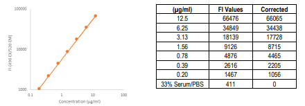
6. Subtract the FI blank values (33% normal mouse plasma in 0.05M phosphate buffered
saline) from the FI values of the standards and samples.
7. Plot the FI values of the standards against the μg/ml of the FITC-Dextran standards. Using
a log/log plot will linearize the data (Figure 1).
Figure 1. A Typical Standard Curve for FITC-Dextran
Related Products
Related Products

FITC-Dextran, 4 kDa, 25 mg/ml x 5 ml

TRITC-Dextran, 70 kDa, 25 mg/ml x 5 ml

AbraMag Biotin Magnetic Beads, 5 mL, 5 mg/mL

AbraMag Streptavidin Magnetic Beads, 5 mL, 5 mg/mL
