
Exploring PDB Structure 9MWM and Selective Inhibitor Design
STRUCTURAL BIOLOGY • ENZYMOLOGY • DRUG DISCOVERY
Neuronal Nitric Oxide Synthase (nNOS): Exploring PDB Structure 9MWM and Selective Inhibitor Design
Discover how X-ray crystallography reveals the interactions between neuronal nitric oxide synthase and a small-molecule inhibitor, supporting research into selective drug discovery.
Understanding the three-dimensional structure of a protein can help researchers explain its biological activity and identify molecules that may alter its function. The Protein Data Bank (PDB) entry 9MWM provides a structural view of the heme domain of rat neuronal nitric oxide synthase (nNOS), an enzyme involved in nitric oxide production. The structure contains an R349A mutant protein domain bound to a small-molecule inhibitor investigated in research on melanoma.
This structure is associated with research into structure-based inhibitor design, where detailed protein–ligand interactions guide the development of molecules with improved potency and selectivity. Explore the 9MWM entry in the RCSB Protein Data Bank.
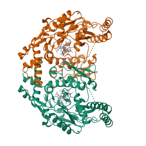
Structure image: RCSB Protein Data Bank, entry 9MWM. Image source: RCSB PDB 9MWM.
1. What Is Neuronal Nitric Oxide Synthase?
Neuronal nitric oxide synthase, commonly abbreviated as nNOS or NOS1, is an enzyme that produces nitric oxide (NO) from L-arginine in a reaction that also requires oxygen and appropriate cofactors. Nitric oxide is a signaling molecule involved in several physiological processes, including communication between nerve cells.
nNOS belongs to the nitric oxide synthase family, which also includes endothelial nitric oxide synthase (eNOS) and inducible nitric oxide synthase (iNOS). Although these enzymes perform related chemical reactions, they have different biological roles and expression patterns.
Because the NOS isoforms share substantial structural similarity, developing an inhibitor that preferentially targets nNOS while minimizing effects on eNOS and iNOS is an important challenge in medicinal chemistry.
2. What Does PDB Entry 9MWM Represent?
PDB 9MWM describes the heme domain of rat neuronal nitric oxide synthase carrying the R349A mutation and bound to a small molecule investigated as an nNOS inhibitor. The ligand is a thiazole-5-carboximidamide derivative with a substituted biphenyl-containing structure.
Structure Information
- PDB ID: 9MWM
- Protein: Neuronal nitric oxide synthase, heme domain
- Organism: Rattus norvegicus (rat)
- Mutation: R349A
- Expression system: Escherichia coli BL21(DE3)
- Experimental method: X-ray diffraction
- Reported resolution: 1.95 Å
- Classification: Oxidoreductase
- Structure DOI: 10.2210/pdb9MWM/pdb
A resolution of 1.95 Å provides detailed information about the modeled atomic arrangement of the protein and bound ligand. This level of structural detail can help scientists examine ligand orientation, binding-site geometry, and potential molecular interactions.
3. Understanding the Heme Domain and Ligand Binding
The heme domain contains a heme group, an iron-containing molecular component involved in the enzyme's catalytic chemistry. The surrounding protein structure helps position substrates and cofactors and creates the molecular environment required for enzyme activity.
In structure-based drug discovery, researchers examine how a candidate inhibitor fits into the enzyme's binding region. Relevant features may include hydrogen bonding, electrostatic interactions, hydrophobic contacts, and the spatial complementarity between the ligand and the protein.
For PDB 9MWM, the structure allows investigators to inspect the bound inhibitor in the context of the rat nNOS heme domain. Structural observations can inform hypotheses about binding, but the biological significance of individual contacts must be evaluated alongside biochemical and functional experiments.
4. Why Selective nNOS Inhibitors Matter
Designing selective inhibitors is a major goal in research on nitric oxide synthases. Inhibitors that bind several NOS isoforms may produce unwanted effects by interfering with physiological functions carried out by other enzymes.
Researchers therefore investigate differences in amino acid residues, binding-site shape, and local chemical environments to identify interactions that favor nNOS over eNOS and iNOS. These differences can be used to guide molecular design and structure–activity relationship (SAR) studies.
The associated 2026 publication, New Inhibitors of Neuronal Nitric Oxide Synthase for the Treatment of Melanoma, describes a structure-based strategy for developing nNOS inhibitors with improved isoform selectivity. The authors report potent inhibition and selectivity for a lead compound in biochemical assays. These findings support further investigation, but they do not establish an approved melanoma treatment.
5. Connection Between nNOS and Melanoma Research
Melanoma is a type of cancer that develops from pigment-producing cells called melanocytes. Research cited in the associated publication investigates the role of nNOS-derived nitric oxide in melanoma progression and explores whether selective inhibition of nNOS could provide a useful therapeutic research direction.
The scientific rationale is to investigate whether modifying this enzyme's activity can influence processes involved in tumor biology. The value of a structural model such as 9MWM is that it helps researchers study the molecular basis of inhibitor binding and refine compounds for subsequent testing.
Importantly, a protein structure alone cannot demonstrate that an inhibitor is safe or effective in patients. Additional biochemical, cellular, animal, and clinical studies are needed to evaluate therapeutic potential.
6. How X-Ray Crystallography Supports Drug Discovery
X-ray crystallography is a structural biology technique used to determine the atomic arrangement of molecules in a crystal. In protein crystallography, researchers collect diffraction data and use computational methods to build and refine a model of the protein and any bound ligands.
For inhibitor research, a protein–ligand structure can help scientists:
- Visualize the orientation of a ligand inside a binding site.
- Identify candidate interactions between the ligand and amino acid residues.
- Compare related inhibitors and understand structure–activity relationships.
- Generate hypotheses for modifying a molecule to improve potency or selectivity.
- Support molecular modeling and future compound-design experiments.
Structural models should be interpreted alongside validation metrics and independent experimental evidence. Resolution, refinement statistics, ligand fit, and local electron-density quality all contribute to evaluating how confidently a molecular interaction can be interpreted.
7. Exploring PDB 9MWM for Research and Education
The 9MWM entry can be used as a reference for studying enzyme structure, ligand recognition, and medicinal chemistry. Researchers and students can explore its three-dimensional coordinates, examine the ligand-binding region, and compare this structure with related NOS protein–ligand complexes.
When analyzing the structure, useful questions include:
- Where is the inhibitor located relative to the heme group?
- Which amino acid residues surround the ligand?
- How does the binding pocket compare with those of other NOS isoforms?
- What additional experiments would help validate the proposed binding interactions?
Answers to these questions can help connect structural observations with enzyme function and inhibitor-design principles.
References and Further Reading
1. RCSB Protein Data Bank. Structure of rat neuronal nitric oxide synthase R349A mutant heme domain bound with a small-molecule inhibitor. PDB entry 9MWM.
2. Awasthi A, Patel A, Li H, et al. New Inhibitors of Neuronal Nitric Oxide Synthase for the Treatment of Melanoma. Journal of Medicinal Chemistry. 2026;69:2310–2329.
Educational content for scientific and research purposes. Experimental findings discussed here should not be interpreted as evidence of an approved treatment or as medical advice.
Recent Posts
-
Recombinant Rabbit Monoclonal Antibodies: Principles, Production and Research Applications
ANTIBODY TECHNOLOGY • MOLECULAR BIOLOGY Recombinant Rabbit Monoclonal Antibodies: Principles, Prod …5th Nov 2026 -
Phagocytosis: Mechanisms, Zymosan-Based Assays and Research Applications
CELL BIOLOGY • IMMUNOLOGY • RESEARCH Phagocytosis: Mechanisms, Zymosan-Based Assays and Research A …5th Nov 2026 -
Exploring PDB Structure 9MWM and Selective Inhibitor Design
STRUCTURAL BIOLOGY • ENZYMOLOGY • DRUG DISCOVERY Neuronal Nitric Oxide Synthase (nNOS): Exploring …29th Oct 2026