
Type I Collagen C-Telopeptide (CTX-I) Assay Kit
 Assay Kit")
- SKU:
- 445-6033
- Size:
- 1 kit
- Shipping:
- Gel Packs
- Storage:
- -20 C
 Assay Kit")
Description
Type I Collagen C-Telopeptide (CTX-I) Assay Kit - Cat Number: 6033 From Chondrex.
Research Field: Arthritis, ECM
Clonality: N/A
Cross-Reactivity:
Host Origin: N/A
Applications: N/A
Isotype: N/A
Detection Range: 500 ng/ml - 7.8 ng/ml
Sample Type: Serum, Plasma, Urine
Concentration: N/A
Immunogen:
PRODUCT SPECIFICATIONS
DESCRIPTION: ELISA Kit to quantify CTX-I fragments/peptides
FORMAT: 96-well ELISA Plate with removeable strips
ASSAY TYPE: Competitive ELISA
ASSAY TIME: 3 hours
STANDARD RANGE: 500 - 8 ng/ml
NUMBER OF SAMPLES: Up to 40 (duplicate) samples/plate
SAMPLE TYPES: Urine, Serum, and Plasma
RECOMMENDED SAMPLE DILUTIONS: 1:100 (at least)
CHROMOGEN: TMB (read at 450 nm)
STORAGE: -20°C for 12 months
VALIDATION DATA: Intra-Assay (1.6-2.2%)/Inter-Assay (5.5-10%)/Spiking Test (108-113%)
INTRODUCTION
Collagen is the most abundant protein in the mammalian body and lends structural integrity to tissues as the primary component of the extracellular matrix (1). Type I collagen is the main component of bone, tendon, skin, and other tissues (2). In fact, type I collagen makes up 20% of bones by mass, which accounts for more than 90% of the organic components. As a result, degradation products of type I collagen can be detected in serum and urine in stages of bone loss or metabolism and can be potential markers of bone metabolism (3). Proteinases mediate resorption of type I collagen from bone and generate specific peptide fragments of degraded collagen. For example, matrix metalloproteinases (MMPs) exclusively produce C-terminal degraded fragments (ICTP) from type I collagen, while cathepsin K produces CTX-I fragments from the C-terminus and NTX-I fragments from the N-terminus of type I collagen (4). Because the proteinase activities differ among diseases and degraded fragments reflect the metabolism of type I collagen, immunoassays have been developed to monitor the levels of these degraded fragments in biological fluids (5).
Patients with osteoporosis display reduced bone mass. During disease progression, degraded peptides of type I collagen are observed in serum as well as urine. Therefore, ICTP and NTX-I have been used as markers of osteoporosis (6-8). In addition, it was reported that CTXI levels in urine correlate with disease activity of osteoarthritis (9). Furthermore, cancers which metastasize to bone can affect the metabolism of type I collagen. Serum CTX-I levels correlate with prognosis of these cancers, especially prostate, lung, breast, and urinary bladder cancers (10-13).
Thus, degraded type I collagen fragments are very useful tools for evaluating disease not only in humans, but also in mice, leading to the development of many mouse disease models for cancer, osteoporosis, osteoarthritis, and rheumatoid arthritis. Chondrex, Inc. has developed a CTX-I Detection ELISA kit (Cat # 6033) for mouse and human samples using a competitive assay system with a monoclonal antibody which recognizes conserved peptide sequences in mouse and human (13).
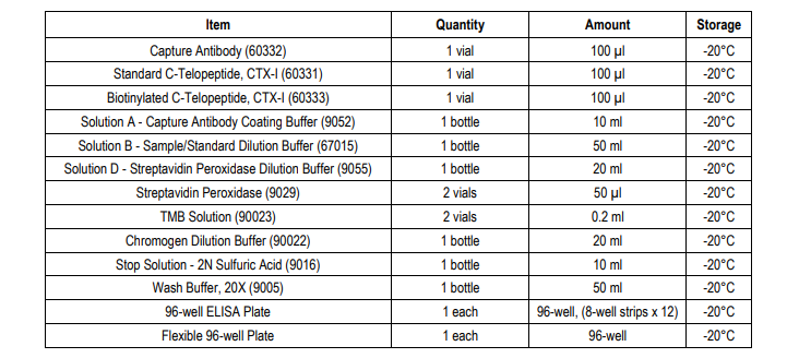
KIT COMPONENTS
ASSAY OUTLINE

NOTES BEFORE USING ASSAY
NOTE 1: It is recommended that the standard and samples be run in duplicate.
NOTE 2: Warm up all buffers to room temperature before use.
NOTE 3: Crystals may form in Wash Buffer, 20X when stored at cold temperatures. If crystals have formed, warm the wash buffer by placing the bottle in warm water until crystals are completely dissolved.
NOTE 4: Measure exact volume of buffers using a serological pipet, as extra buffer is provided.
NOTE 5: Cover the plate with plastic wrap or a plate sealer after each step to prevent evaporation from the outside wells of the plate.
NOTE 6: For partial reagent use, please see the assay protocol’s corresponding step for the appropriate dilution ratio. For example, if the
protocol dilutes 50 µl of a stock solution in 10 ml of buffer for 12 strips, then for 6 strips, dilute 25 µl of the stock solution in 5 ml of buffer.
Partially used stock reagents may be kept in their original vials and stored at -20⁰C for use in a future assay.
NOTE 7: This kit contains animal components from non-infectious animals and should be treated as potential biohazards in use and for disposal.
ASSAY PROCEDURE
1. Add Capture Antibody: Dilute one vial of Capture Antibody with 10 ml of Capture Antibody Dilution Buffer (Solution A). Alternatively, dilute according to table below. Add 100 µl of capture antibody solution to each well and incubate at 4°C overnight. Any leftover Capture Antibody Stock Solution may be stored at -20°C for future assays.
TROUBLESHOOTING
For frequently asked questions about assays and ELISAs, please see Chondrex, Inc.’s ELISA FAQ for more information.
REFERENCES
1. E. Hohenester, J. Engel, Domain structure and organisation in extracellular matrix proteins. Matrix Biol 21, 115-28 (2002).
2. K. Gelse, E. Pöschl, T. Aigner, Collagens--structure, function, and biosynthesis. Adv Drug Deliv Rev 55, 1531-46 (2003).
3. E. Eriksen, K. Brixen, P. Charles, New markers of bone metabolism: clinical use in metabolic bone disease. Eur J Endocrinol 132, 251- 63 (1995).
4. G. Wheater, M. Elshahaly, S. Tuck, H. Datta, L. van, The clinical utility of bone marker measurements in osteoporosis. J Transl Med 11, 201 (2013).
5. J. Risteli, I. Elomaa, S. Niemi, A. Novamo, L. Risteli, Radioimmunoassay for the pyridinoline cross-linked carboxy-terminal telopeptide of type I collagen: a new serum marker of bone collagen degradation. Clin Chem 39, 635-40 (1993).
6. M. Bonde, C. Fledelius, P. Qvist, C. Christiansen, Coated-tube radioimmunoassay for C-telopeptides of type I collagen to assess bone resorption. Clin Chem 42, 1639-44 (1996).
7. K. Lee, M. Lee, C. Chung, W. Seong, S. Lee, M. Park, et al., Measurement of urinary N-telopeptides and serum C-telopeptides from type I collagen using a lateral flow-based immunoassay. Sensors (Basel) 13, 165-74 (2012).
8. J. Clemens, M. Herrick, F. Singer, D. Eyre, Evidence that serum NTx (collagen-type I N-telopeptides) can act as an immunochemical marker of bone resorption. Clin Chem 43, 2058-63 (1997).
9. S. Ok, S. Lee, H. Park, S. Jeong, C. Ko, Y. Kim, et al., Concentrations of CTX I, CTX II, DPD, and PYD in the urine as a biomarker for the diagnosis of temporomandibular joint osteoarthritis: A preliminary study. Cranio 36, 366 372 (2018).
10. A. Ferreira, I. Alho, S. Casimiro, L. Costa, Bone remodeling markers and bone metastases: From cancer research to clinical implications. Bonekey Rep 4, 668 (2015).
11. A. Zissimopoulos, K. Stellos, D. Matthaios, G. Petrakis, V. Parmenopoulou, et al., Type I collagen biomarkers in the diagnosis of bone metastases in breast cancer, lung cancer, urinary bladder cancer and prostate cancer. Comparison to CEA, CA 15-3, PSA and bone scintigraphy. J BUON 14, 463-72 (Jul-).
12. A. Franjević, R. Pavićević, G. Bubanović, ICTP in bone metastases of lung cancer. Coll Antropol. 35, 43–47 (2011).
13. A. Srivastava, S. Bhattacharyya, G. Castillo, N. Miyakoshi, S. Mohan, D. Baylink, et al., Development and evaluation of C-telopeptide enzyme-linked immunoassay for measurement of Bone resorption in mouse serum. Bone 27, 529-33 (2000).
Related Products
Related Products
 Detection Kit")
Type I Collagen N-Telopeptide (NTX-I) Detection Kit

Rapid Collagenase Assay Kit with Type I Collagen Substrate

Mouse Anti-Bovine Type I Collagen IgG Antibody Assay Kit, OPD

Mouse Anti-Porcine Type I Collagen IgG Antibody Assay Kit, TMB
