
Alexa Fluor® 488 (AF488) Conjugated anti-SAH Monoclonal Antibody Clone 839-6 | MAF00302-50

- SKU:
- ART-MAF00302-50
- Availability:
- Usually Shipped in 5 Working Days
- Size:
- 50 µg

Description
Alexa Fluor® 488 (AF488) Conjugated anti-SAH Monoclonal Antibody Clone 839-6 | MAF00302-50 | Arthus Biosystems
Product name
Alexa Fluor 488-anti-SAH 2
Catalog Number
MAF00302-50
Description
Alexa Fluor® 488 (AF488) conjugated anti-S-adenosylhomocysteine monoclonal antibody clone 839-6
Specificity
MAF00302 shows the same specificity as un-conjugated mouse anti-SAH monoclonal antibody MA00307.
Properties
Form
Liquid
Storage instructions
Store at 2-8°C in dark, do not freeze.
Concentration
2-4ring/m1 or lot specific
Storage buffer
50mM Tris, 150mM NaCI, pH8.0, 0.2% BSA (Sigma), 0.09%NaN3
Dilution buffer
PBS, pH 7.4, 1% fetal bovine serum or 0.5% BSA, 0.09%NaN3
Purity
>95% purified with Sephadex G-25, free from un-conjugated antibody and Alexa Fluor 488
Clonality
Monoclonal
Clone number
839-6
Immunoglobin isotype
mouse IgG2a
Research Areas
- Methylation of biomolecules (DNA, RNA, proteins, hormones, neurotransmitters, etc.)
- One-carbon metabolism
- Signal Transduction
- Metabolism
- Pathways and Processes Cancer
- Arthritis
- Neurodegenerative diseases Atherosclerosis
- Liver diseases
- Kidney diseases
Applications
The use of MAF00302 in the following applications has been tested. The application notes include recommended and tested dilutions. Optimal dilutions/concentrations should be determined by the end user based on the test environment and purposes.
Recommended
Flow Cytometry (FCM): 20-80 pg/ml
Immunofluorescence Laser Scanning Confocal Microscopy (LSCM): 30-60 pg/ml
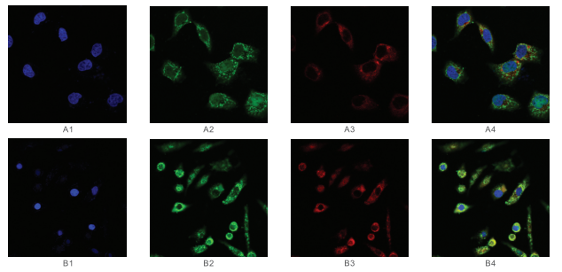
Figure 1 : Immunofluorescence (IF) LSCM results of normal liver cells L02 cultured in RPMI 1640 with 10% FBS for 16h followed by stimulation by 0.5mM methionine for 24h (Al -A4) double stained with AF488-anti-SAH 839-6 (Cat# MAF00302) at 40pg/m1 and R-PE-anti-SAM 84-3 (Cat# MAF00202) at 40pg/m1 followed by DAPI staining. Hepatocellular carcinoma cell line HepG2 cells were cultured in RPMI 1640 with 10% FBS for 16h followed by stimulation by 0.5mM methionine for 24h (B1-B4) and double stained with AF488-anti-SAH 839-6 (Cat# MAF00302) at 60pg/m1 and R-PE-anti-SAM 84-3 (Cat# MAF00202) at 60pg/m1 followed by DAPI staining. Photography was performed under the laser scanning confocal microscope Zeiss LSM 780 (x630). Different views are as follows: DAPI (Al, B1); AF488 for SAH (A2, B2); R-PE for SAM (A3, B3); Overlap of all the three fluorescent signals (A4, B4). Expression patterns of SAM and SAH are different between L02 and HepG2 cells after methionine stimulation for 24h. In the case of cells not actively proliferating, both SAM and SAH are seen more in cytoplasm (more in mitochondria areas) than nuclear.
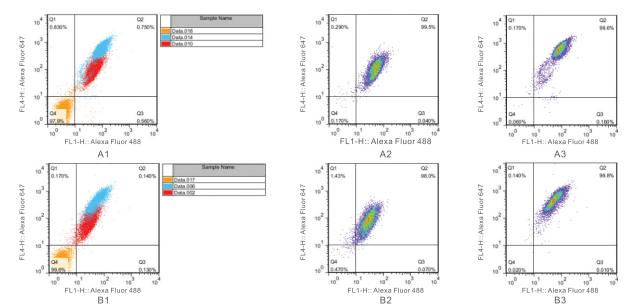
Figure 2: Flow Cytometry of L02 (Al -A3) and HepG2 (B1-B3) cells double stained with Alexa Fluor® 488 conjugated anti-SAH antibody 839-6 (Cat# MAF00302) at 18 pg/ml and Alexa Fluor®647 conjugated anti-SAM antibody 118-6 (Cat# MAF00201) at 4.5 pg/ml. Color legend: Orange: blank; Blue: nuclear fixation/permeabilization buffer was used (eBioscience 00-5523 FoxP3_TF Staining Buffer Set); Red: intracellular fixation/permeabilization buffer was used (eBioscience 00-8824). 100% confluent cells (cultured for 48h) were fixed and permeabilized with the intracellular fixation/permeabilization buffer (A2, B2) or the nuclear fixation/permeabilization buffer (A3, B3) and then double stained with antibodies indicated above. Cells were used for analysis with BD FACSCalibur Flow Cytometer. SAM expression is higher in L02 than HepG2 cells. Both SAM and SAH are expressed ubiquitously yet rather dynamically. The level of SAM is higher than that of SAH in both cells.
1 Review
-
Quick shipping
Gentaur always fulfils its promise. They always shipped their products on time without any delay. Highly recommended!
Related Products
Related Products

Alexa Fluor® 488 (AF488) Conjugated anti-SAH Monoclonal Antibody Clone 301-3 | MAF00301

Alexa Fluor® 647 (AF6) Conjugated anti-SAM Monoclonal Antibody Clone 118-6 | MAF00201
![AF488-streptavidin conjugate [Streptavidin, Alexa Fluor(tm) 488 Conjugate] | 16891](https://cdn11.bigcommerce.com/s-ryt90hjx0j/images/stencil/590x590/products/36268/45875/Capture_dcran_2022-08-10_182314__97365.1660152248.png?c=1 "AF488-streptavidin conjugate [Streptavidin, Alexa Fluor(tm) 488 Conjugate] | 16891")
AF488-streptavidin conjugate [Streptavidin, Alexa Fluor(tm) 488 Conjugate] | 16891

HRP Anti-SAH Monoclonal Antibody Clone 301-16 | MAH00302-50
