
DNA Assay Kit

- SKU:
- 445-6023
- Size:
- 1 kit
- Shipping:
- Gel Packs
- Storage:
- -20 C

Description
DNA Assay Kit - Cat Number: 6023 From Chondrex.
Research Field: SLE
Clonality: N/A
Cross-Reactivity:
Host Origin: N/A
Applications: N/A
Isotype: N/A
Detection Range: 10 µg/ml-0.16 µg/ml
Sample Type: Tissue Extract, Cultured Cells
Concentration: N/A
Immunogen:
PRODUCT SPECIFICATIONS
DESCRIPTION: Assay kit to quantify DNA
FORMAT: 96-well ELISA plate with non-removeable strips
ASSAY TYPE: Dye-based assay/Fluorescence-based assay
ASSAY TIME: <0.5 hour
STANDARD RANGE: 10 µg/ml to 0.16 µg/ml
NUMBER OF SAMPLES: Up to 40 (duplicate) samples/plate
SAMPLE TYPES: Solubilized tissue & cell culture samples (pre-treatment acceptable)
RECOMMENDED SAMPLE DILUTIONS: Varies (Typical DNA levels are usually 0.3-5 µg/mg wet tissue and 1-5 µg/1x106 cells)
FLUORESCENCE: Excitation 360 nm/Emission 460 nm
STORAGE: -20°C for 12 months
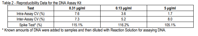
VALIDATION DATA: Intra-Assay (1.7-7.6%)/Inter-Assay (5.2-8.0%)/Spiking Test (105.1-116.2%)
INTRODUCTION
Deoxyribonucleic acid (DNA) is a distinct cell component, therefore DNA amount is correlated with cell number (1). For example, in cartilage tissue engineering, artificial cartilage quality is evaluated by DNA amounts translated as chondrocyte numbers, as well as amounts of collagen and glycosaminoglycans (GAGs) in extracellular matrix components (ECM) (2, 3). DNA can be quantified by the ratio of absorbance at 260 nm and 280 nm, but this method is affected by contaminating proteins, RNA, and chemicals in the sample, requiring additional DNA isolation steps. For tissue analysis, Chondrex, Inc. provides an alternative DNA assay kit employing the Hoechst 33258 fluorescent dye which specifically binds to Adenine-Thymine base pairs, resulting in fluorescence at excitation 360 nm/emission 460 nm. Because the dyeDNA binding and the fluorescence intensity are unaffected by contaminating proteins and other substances in an optimized assay condition, this DNA assay kit works accurately with samples containing other analytes. In order to facilitate further studies in this field, Chondrex, Inc. also provides various native collagen detection ELISAs, a Hydroxyproline. Assay Kit (Cat # 6017), Sirius Red/ Fast Green Staining Kit (Cat # 9046 ), Sirius Red Total Collagen Detection Kit (Cat # 9062), and GAGs assay kit (Cat # 6022). Please visit www.chondrex.com or contact support@chondrex.com for more information.
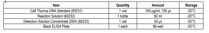
KIT COMPONENTS
ASSAY OUTLINE
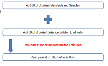
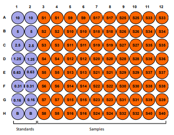
PLATE MAPPING
NOTES BEFORE USING ASSAY
NOTE 1: It is recommended that the standard and samples be run in duplicate.
NOTE 2: Warm up all buffers to room temperature before use.
NOTE 3: Crystals may form in Wash Buffer, 20X when stored at cold temperatures. If crystals have formed, warm the wash buffer by placing
the bottle in warm water until crystals are completely dissolved.
NOTE 4: Measure exact volume of buffers using a serological pipet, as extra buffer is provided.
NOTE 5: Cover the plate with plastic wrap or a plate sealer after each step to prevent evaporation from the outside wells of the plate.
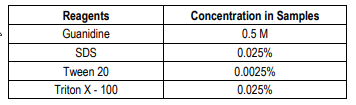
Table 1 - Solubilizing Reagents in the DNA Assay Kit
ASSAY PROCEDURE
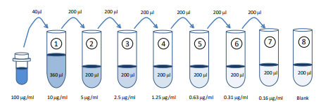
1. Prepare Standard Dilutions: Take 40 µl of DNA Standard Solution and mix with 360 µl of Reaction Solution (10 µg/ml). Then serially
dilute it with Reaction Solution. For example, mix 200 µl of the standard (10 µg/ml) with an equal volume of Reaction Solution to make
a 5 µg/ml solution, and then repeat it five more times for 2.5, 1.3, 0.63 0.31, and 0.16 µg/ml solutions. Chondrex, Inc. recommends
making fresh serial dilutions for each assay.
2. Prepare Sample Dilutions: Dilute samples with Reaction Solution. The optimal sample dilution depends on the sample and preparation
methods; therefore, Chondrex, Inc. recommends multiple dilutions as a preliminary study.
3. Prepare 1X Detection Solution: Mix 5 µl of 200X Concentrated Detection Solution and 1 ml Reaction Solution for each well. For
example, 8 samples, 7-point standards, one blank (all in duplicate) will require 1.6 ml of the 1X Detection Solution. Mix 8 µl of 200X
Concentrated Detection Solution and 1.6 ml Reaction Solution.
NOTE: Prepare the 1X solution just before use. Do not store and reuse the mixed solution for the next assay.
4. Add Standards and Samples: Add 50 µl of diluted standards and samples into wells.
5. Add 1X Detection Solution: Add 50 µl of 1X detection solution into all wells, and then incubate at room temperature for 5 minutes.
6. Read plate: Read the plate at excitation 360 nm/emission 460 nm.
CALCULATING RESULTS
1. Average the duplicate fluorescence intensity (FI) values for the blank, standards, and test samples.
2. Subtract the averaged “blank” (B) FI values from the averaged FI values of standards and test samples.
3. Plot the FI values of the standards against the DNA concentration (µg/ml). Using a log/log plot will linearize the data. Figure 1 shows
an example of a DNA standard curve from 0.16-10 µg/ml.
4. The μg/ml of DNA in test samples can be calculated using regression analysis. Multiply it by the sample dilution factor to obtain the
DNA concentration (μg/ml) in the original sample.
NOTE 1: DNA levels in samples depend on the sample solubilization efficiency of individual protocols consequently, converting DNA
levels to cell numbers is not always applicable. Therefore, DNA levels as correlating to cell numbers can be used to compare samples
including positive and negative controls.
NOTE 2: To correlate DNA amounts to cell numbers, validating your DNA solubilization protocol is required. Please refer to the
references for more information.

ASSAY VALIDATION
NOTES:
Related Products
Related Products

Glycosaminoglycans Assay Kit

Hydroxyproline Assay Kit

Creatinine Assay Kit

Hemoglobin Assay Kit
